
Pain reflex arcs and the cerebellumSpec C3.1.9, C3.1.10
In short
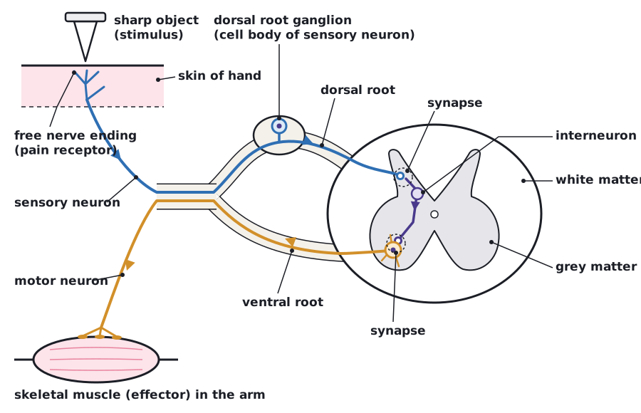
A pain reflex arc is an involuntary response with skeletal muscle as the effector. A free nerve ending of a sensory neuron in the hand detects pain, the impulse passes to a single interneuron in the grey matter of the spinal cord, then to a motor neuron that makes a muscle contract. The cerebellum coordinates skeletal muscle contraction and balance.
A reflex is a rapid, involuntary response to a stimulus. The pain reflex arc is an example of an involuntary response with skeletal muscle as the effector. Because the spinal cord, not the brain, processes the signal, the response is fast and does not need a conscious decision.
- A sharp object pricks the hand. A free sensory nerve ending of a sensory neuron acts as the pain receptor and generates nerve impulses.
- The sensory neuron carries impulses to the spinal cord, entering through the dorsal root.
- In the grey matter of the spinal cord, the sensory neuron synapses with a single interneuron.
- The interneuron synapses with a motor neuron, which carries impulses out of the spinal cord through the ventral root.
- The motor neuron stimulates a skeletal muscle (for example the biceps) to contract, pulling the hand away.
Other interneurons also pass impulses up the spinal cord to the brain, so the pain is felt consciously, but only after the withdrawal has already begun.
 (opens full size in a new tab)
The cerebellum
The cerebellum, at the back of the brain below the cerebral hemispheres, coordinates skeletal muscle contraction and balance. It receives information about body position from muscles, joints, the inner ear and the eyes, and adjusts the timing and strength of muscle contractions so that movements are smooth and accurate and posture is maintained. It is important in learned motor skills such as walking, riding a bicycle or writing.
The cerebellum does not start voluntary movements; the cerebral hemispheres do. The cerebellum coordinates them and keeps the body balanced.
Written and checked against the IB Biology HL specification · Updated October 2026