
Single and double circulation and the mammalian heartSpec B3.2.14, B3.2.15
In short
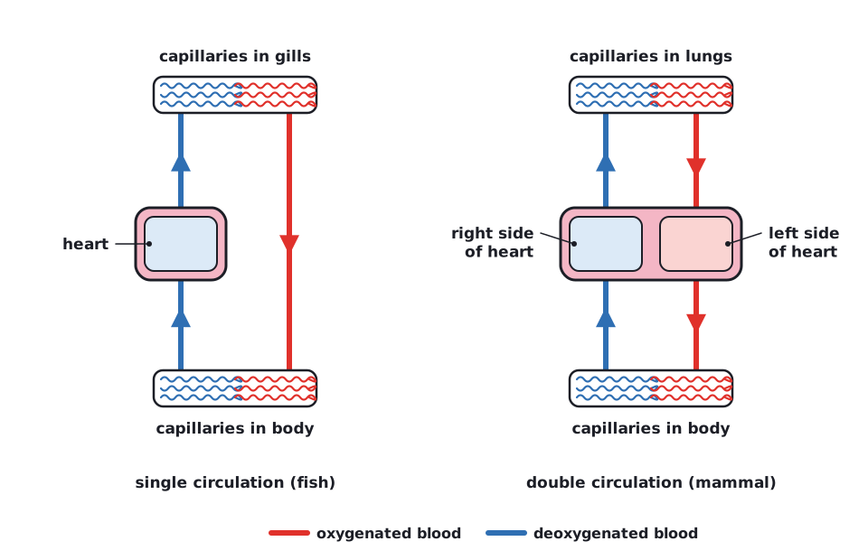
Bony fish have a single circulation: blood passes through the heart once, from heart to gills to body. Mammals have a double circulation: blood passes through the heart twice, through separate pulmonary and systemic circuits. The mammalian heart delivers pressurized blood using myogenic cardiac muscle, a pacemaker, thick-walled ventricles, valves, a septum and coronary vessels.
 (opens full size in a new tab)
| Feature | Bony fish (single) | Mammal (double) |
|---|---|---|
| Sequence of organs | Heart → gills → body → heart | Heart (right side) → lungs → heart (left side) → body → heart |
| Passes through heart per circuit | Once | Twice |
| Pressure of blood reaching the body | Low, because pressure is lost in the gill capillaries | High, because blood is pumped again after the lungs |
| Separation of blood | Only deoxygenated blood in the heart | Septum keeps oxygenated and deoxygenated blood apart |
Adaptations of the mammalian heart
| Structure | Adaptation |
|---|---|
| Cardiac muscle | Contracts without nerve stimulation (myogenic); branched, connected cells pass impulses rapidly so the walls contract together; does not fatigue. |
| Pacemaker (sinoatrial node) | A group of specialised cells in the wall of the right atrium that initiates each heartbeat. |
| Atria | Thin-walled chambers that collect blood from veins and pump it a short distance into the ventricles. |
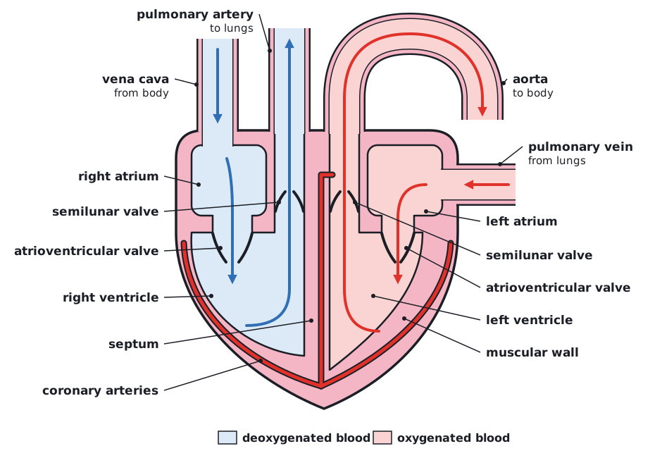
| Ventricles | Thick muscular walls generate high pressure; the left ventricle wall is thicker because it pumps blood around the whole body. |
| Atrioventricular valves | Close when ventricles contract, preventing backflow into the atria. |
| Semilunar valves | In the aorta and pulmonary artery; close when ventricles relax, preventing backflow into the ventricles. |
| Septum | Wall separating left and right sides, keeping oxygenated and deoxygenated blood apart. |
| Coronary vessels | Supply cardiac muscle with oxygen and glucose and remove CO₂. |
 (opens full size in a new tab)
- Vena cava → right atrium → atrioventricular valve → right ventricle → semilunar valve → pulmonary artery → lungs.
- Pulmonary veins → left atrium → atrioventricular valve → left ventricle → semilunar valve → aorta → body.
Written and checked against the IB Biology HL specification · Updated October 2026