
Synapses and excitatory postsynaptic potentialsSpec C2.2.5, C2.2.6, C2.2.7
In short
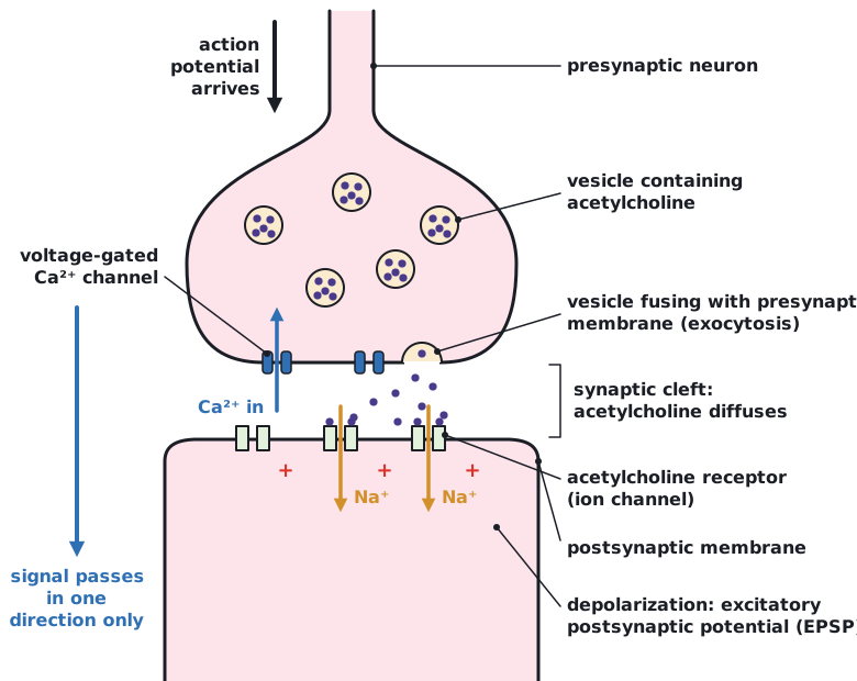
A synapse is a junction between two neurons or between a neuron and an effector cell. Depolarization of the presynaptic membrane lets calcium ions in, which triggers release of neurotransmitter by exocytosis. Acetylcholine diffuses across the synaptic cleft, binds to transmembrane receptors and lets positive ions in, generating an excitatory postsynaptic potential.
Synapses are junctions between neurons and between neurons and effector cells, such as muscle fibres at neuromuscular junctions. At a chemical synapse (called simply a synapse here) the cells are separated by a narrow synaptic cleft.
A signal can only pass in one direction across a typical synapse. Only the presynaptic neuron has vesicles of neurotransmitter, and only the postsynaptic membrane has the receptors.
Release of neurotransmitter from the presynaptic membrane
- An action potential arrives and depolarizes the presynaptic membrane.
- Voltage-gated calcium channels open and Ca²⁺ diffuses into the presynaptic neuron (it is more concentrated outside).
- Inside the neuron, calcium ions act as a signalling chemical: they cause vesicles of neurotransmitter to move to the presynaptic membrane and fuse with it.
- The neurotransmitter is released into the synaptic cleft by exocytosis.
Excitatory postsynaptic potential (EPSP)
- Acetylcholine diffuses across the synaptic cleft.
- It binds to transmembrane receptors in the postsynaptic membrane.
- The receptors open ion channels and positively charged ions, mainly Na⁺, diffuse into the postsynaptic cell.
- The postsynaptic membrane is depolarized: an excitatory postsynaptic potential. If it reaches the threshold potential, an action potential starts in the postsynaptic cell.
- The neurotransmitter is quickly removed from the synaptic cleft, so each signal is brief.
Beyond the syllabus: the guide does not name the enzyme that removes acetylcholine. It is acetylcholinesterase, which breaks acetylcholine into acetate and choline; the presynaptic neuron reabsorbs the choline to make more acetylcholine.
Acetylcholine is used at many types of synapse, including neuromuscular junctions, where it triggers contraction of skeletal muscle fibres.
 (opens full size in a new tab)
Linking question: in what ways are biological systems regulated? Synapses are control points: whether a signal passes depends on how much neurotransmitter is released and how many receptors respond.
Written and checked against the IB Biology HL specification · Updated October 2026