
Phases of mitosisSpec D2.1.7, D2.1.8
In short
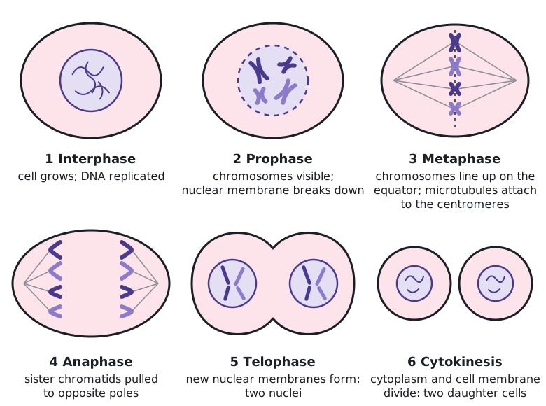
Mitosis has four phases: prophase, metaphase, anaphase and telophase. Chromosomes condense, line up on the equator, have their sister chromatids pulled to opposite poles, and are enclosed in two new nuclei. Because each nucleus receives one copy of every chromatid, mitosis produces two genetically identical daughter nuclei.
- Prophase: chromosomes condense and become visible, each as two sister chromatids. Spindle microtubules start to form, and the nuclear membrane breaks down at the end of prophase.
- Metaphase: microtubules attach to the centromeres. Chromosomes are lined up in a single line on the equator of the cell.
- Anaphase: the centromeres split, so the sister chromatids separate. Each chromatid is now a chromosome, and the two sets are pulled to opposite poles.
- Telophase: chromosomes reach the poles and uncoil. A nuclear membrane forms around each set, giving two nuclei. Cytokinesis usually starts.
The process as a whole gives two genetically identical daughter nuclei. DNA replication made an exact copy of every chromosome, and anaphase sends one chromatid of every chromosome to each pole, so each nucleus receives a complete, identical set.
 (opens full size in a new tab)
Identifying the phases
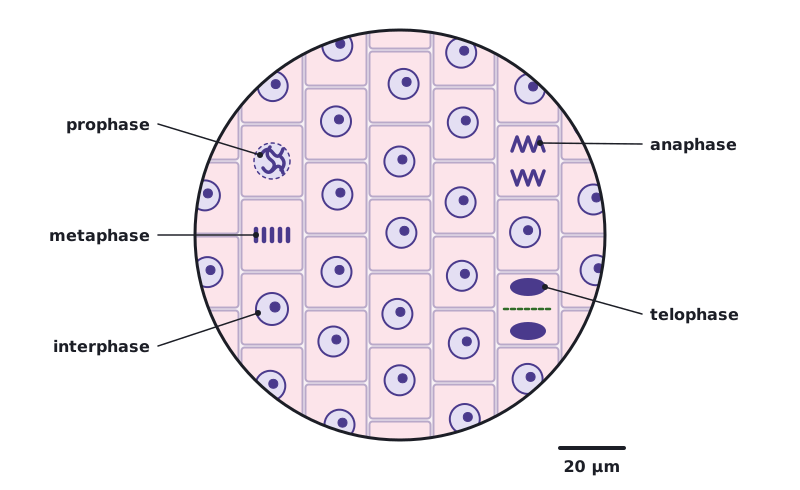
| Phase | Key features you can see |
|---|---|
| Interphase | Intact nucleus, often with a nucleolus; chromosomes not visible as separate threads |
| Prophase | Chromosomes visible as thick threads inside the nuclear area; no metaphase plate yet |
| Metaphase | Chromosomes in a line across the middle of the cell; no nuclear membrane |
| Anaphase | Two groups of V-shaped chromosomes moving apart towards opposite poles |
| Telophase | Two compact groups of chromosomes at the poles; nuclear membranes re-forming; a cell plate or furrow may be visible |
Root tip squashes (for example onion or garlic) show many dividing cells. The tip is softened in warm dilute hydrochloric acid, stained (for example with toluidine blue or acetic orcein) and squashed under a cover slip so cells form a single layer for viewing with a light microscope.
 (opens full size in a new tab)
In anaphase, chromatids move with the centromere first, so the arms trail behind in a V shape. That V shape is the quickest way to spot anaphase in a micrograph.
Written and checked against the IB Biology HL specification · Updated October 2026