
The nucleus, free ribosomes and rough ERSpec B2.2.6, B2.2.7
In short
The nucleus is surrounded by a double membrane, the nuclear envelope, with pores that let mRNA out and proteins in, and it breaks into vesicles during mitosis and meiosis. Free ribosomes make proteins kept inside the cell, while ribosomes bound to the rough endoplasmic reticulum make proteins for transport within the cell or secretion.
The double nuclear membrane
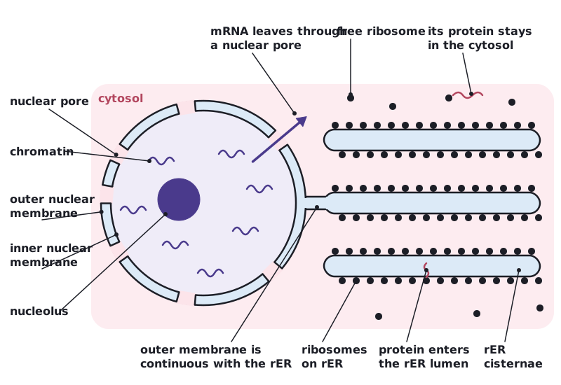
The nucleus is enclosed by two membranes, together called the nuclear envelope. The outer membrane is continuous with the rough endoplasmic reticulum.
- Separation: DNA and transcription are kept apart from translation and other reactions in the cytoplasm.
- Nuclear pores: mRNA made by transcription must leave the nucleus, and proteins made in the cytoplasm (such as enzymes for DNA replication and histones) must enter. Pores, where the two membranes join, allow and control this traffic.
- Breakdown into vesicles: during mitosis and meiosis the nuclear membrane breaks into vesicles so that spindle microtubules can reach and move the chromosomes. Afterwards the vesicles fuse to form new nuclear envelopes around each set of chromosomes.
Free ribosomes and rough ER
Ribosomes are made of rRNA and protein and have no membrane. In eukaryotic cells they are found free in the cytosol or bound to the rough endoplasmic reticulum (rER), a network of flattened membrane sacs called cisternae.
| Feature | Free ribosomes | Ribosomes on the rER |
|---|---|---|
| Location | In the cytosol, not attached to membranes | Attached to the cytoplasmic surface of the rER |
| Proteins made | Proteins for retention in the cell, used in the cytoplasm or in organelles such as mitochondria and the nucleus | Proteins for transport within the cell (for example to lysosomes) or for secretion |
| Where the polypeptide goes | Released into the cytosol | Into the lumen of the rER, then in vesicles to the Golgi apparatus |
Free and rER-bound ribosomes have the same structure, and translation always begins on a free ribosome in the cytosol. If the polypeptide being made begins with a signal sequence (a short stretch of mainly hydrophobic amino acids), the ribosome is guided to the rER and attaches to it. The growing polypeptide is fed through a pore in the rER membrane into the lumen, and the signal sequence is usually cut off. The details of this targeting are not required.
 (opens full size in a new tab)
Written and checked against the IB Biology HL specification · Updated October 2026