
Origin of eukaryotic cells by endosymbiosisSpec A2.2.12
In short
Endosymbiosis is the theory that mitochondria and chloroplasts evolved from free-living prokaryotes that were taken into another cell and survived inside it. All eukaryotes evolved from a common unicellular ancestor with a nucleus; mitochondria arose first, then chloroplasts in some lineages. Evidence includes their 70S ribosomes, naked circular DNA and ability to replicate.
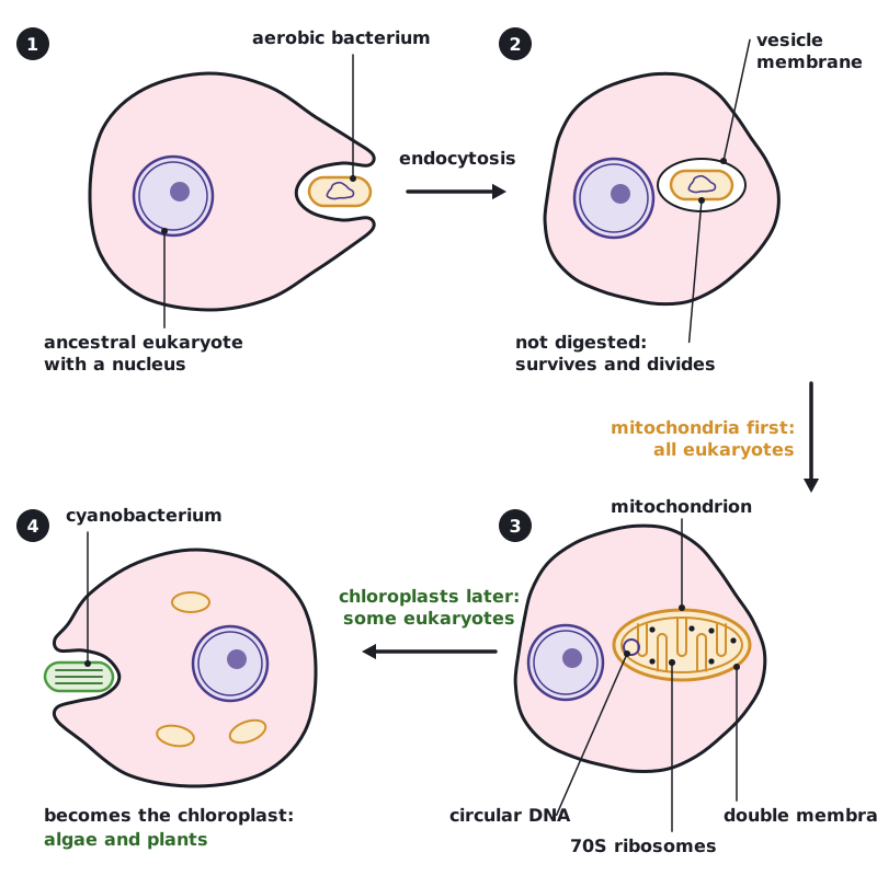
Evidence suggests that all eukaryotes evolved from a common unicellular ancestor that had a nucleus and reproduced sexually. Mitochondria and chloroplasts then arose by endosymbiosis.
- An ancestral eukaryotic cell took in an aerobic prokaryote by endocytosis, but did not digest it.
- The prokaryote survived and divided inside the host. The host gained ATP from the prokaryote's aerobic respiration, and the prokaryote gained protection and a supply of carbon compounds.
- Over many generations the two became dependent on each other, and the prokaryote evolved into the mitochondrion. All eukaryotes are descended from this cell.
- Later, in some eukaryotes only, a photosynthetic prokaryote (a cyanobacterium) was taken in the same way and became the chloroplast, giving rise to algae and plants.
| Evidence | What it shows |
|---|---|
| 70S ribosomes | The same size as prokaryote ribosomes, not the 80S ribosomes of the eukaryotic cytoplasm |
| Naked circular DNA | Like a prokaryote chromosome, not linear chromosomes bound to histones |
| Ability to replicate | They divide by a process like binary fission; a cell cannot make them from scratch |
| Double membrane | Consistent with a prokaryote being taken in by a host cell; the inner membrane resembles a prokaryote plasma membrane |
| Gene base sequences | Most similar to those of aerobic bacteria (mitochondria) and cyanobacteria (chloroplasts) |
 (opens full size in a new tab)
Nature of science: a theory is strong when it explains many observations. Endosymbiosis accounts for the ribosomes, DNA, membranes, division and gene sequences of both organelles.
Written and checked against the IB Biology HL specification · Updated October 2026