
Processes of life in unicellular organismsSpec A2.2.7
In short
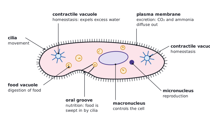
A unicellular organism must carry out all the functions of life within one cell: homeostasis, metabolism, nutrition, movement, excretion, growth, response to stimuli and reproduction. In Paramecium, for example, contractile vacuoles expel excess water for homeostasis, cilia provide movement, food vacuoles digest food, and the cell reproduces by dividing in two.
A unicellular organism consists of one cell, so that one cell must carry out all the functions of life.
- Homeostasis
- Keeping internal conditions within narrow limits
- Metabolism
- All the enzyme-catalysed reactions in the cell
- Excretion
- Removal of the waste products of metabolism
| Function | Paramecium (heterotroph) | Chlamydomonas (autotroph) |
|---|---|---|
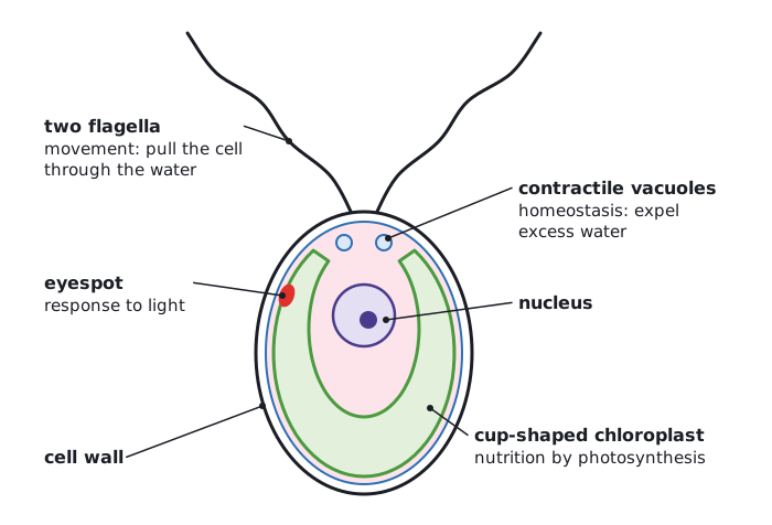
| Homeostasis | Contractile vacuoles collect and expel excess water that enters by osmosis | Contractile vacuoles expel excess water |
| Metabolism | Enzyme-catalysed reactions in the cytoplasm and mitochondria, including respiration | Reactions in the cytoplasm, mitochondria and chloroplast, including photosynthesis and respiration |
| Nutrition | Ingests bacteria and other food into food vacuoles, where it is digested | Makes its own food by photosynthesis in a large cup-shaped chloroplast |
| Movement | Beating cilia over the cell surface | Two flagella pull the cell through the water |
| Excretion | Wastes such as carbon dioxide and ammonia diffuse out across the plasma membrane | Oxygen from photosynthesis and other wastes diffuse out |
| Growth | Increases in size and dry mass by making new molecules | Increases in size and dry mass by making new molecules |
| Response to stimuli | Reverses direction when it meets an obstacle or harmful chemical | An eyespot detects light, so it swims towards light |
| Reproduction | Asexually, by dividing in two (binary fission) | Asexually by mitosis, or sexually when gametes fuse |
 (opens full size in a new tab)
 (opens full size in a new tab)
Learn the eight functions as a list: homeostasis, metabolism, nutrition, movement, excretion, growth, response to stimuli and reproduction. Questions often ask how a named organism carries out each one.
Written and checked against the IB Biology HL specification · Updated October 2026