
Animal, fungal and plant cells, and atypical cellsSpec A2.2.8, A2.2.9
In short
Plant, fungal and animal cells differ in their cell walls, vacuoles, plastids, centrioles, cilia and flagella. Plants have cellulose walls, large permanent vacuoles and chloroplasts; fungi have chitin walls and no plastids; animals have no wall but have centrioles. Atypical cells include multinucleate aseptate fungal hyphae and skeletal muscle fibres, and red blood cells and sieve tubes without nuclei.
| Feature | Animal cells | Fungal cells | Plant cells |
|---|---|---|---|
| Cell wall | Absent | Present, made of chitin | Present, made of cellulose |
| Vacuoles | Small and temporary, such as food vacuoles and vesicles | Present; can be large in older hyphae | A large permanent central vacuole of cell sap; maintains turgor and stores substances |
| Chloroplasts and other plastids | Absent | Absent | Chloroplasts in green tissues; other plastids, such as amyloplasts, store starch |
| Centrioles | Present | Absent in most fungi | Absent in flowering plants |
| Cilia and flagella | Present in some cells, such as ciliated epithelium and the sperm flagellum | Absent in most fungi; only some produce spores with flagella | Absent in flowering plants; mosses and ferns have sperm with flagella |
Common mistake:
Not every plant cell has chloroplasts: root cells have none. Write 'plant cells may have chloroplasts' when comparing cell types.
Atypical cell structure in eukaryotes
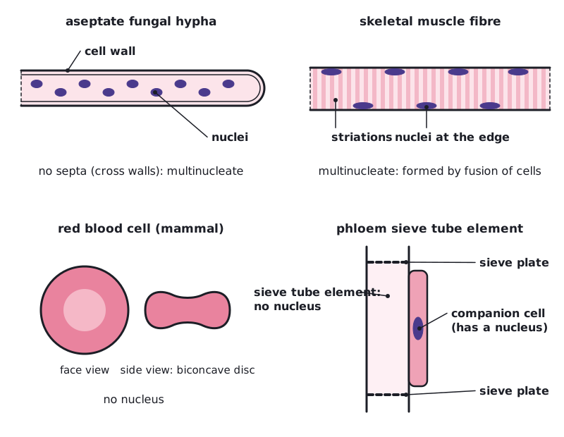
Cell theory pictures a cell as one unit with one nucleus. The numbers of nuclei show that some eukaryotic cells are atypical.
| Cell | Number of nuclei | Explanation |
|---|---|---|
| Aseptate fungal hyphae | Many | The long hyphae have no cross walls (septa), so many nuclei share one continuous cytoplasm |
| Skeletal muscle fibres | Many | Each fibre forms when many embryonic muscle cells fuse, and it can be several centimetres long |
| Red blood cells (mammals) | None | The nucleus is lost as the cell matures, leaving more space for haemoglobin; the cell cannot divide and lives about 120 days |
| Phloem sieve tube elements | None | The nucleus and most organelles break down so sap can flow freely; a companion cell with a nucleus keeps each element alive |
 (opens full size in a new tab)
Written and checked against the IB Biology HL specification · Updated October 2026