
Changes in blood supply to organs with activitySpec D3.3.11
In short
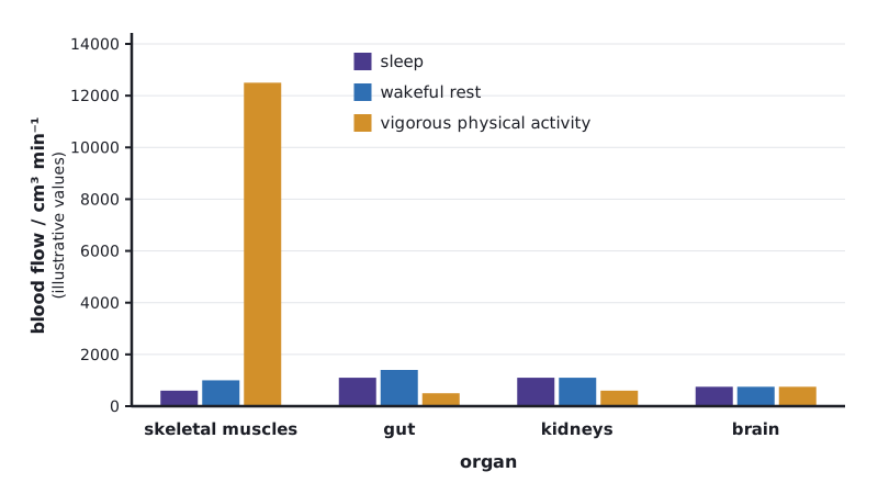
The blood supply to organs changes with activity because arterioles dilate or constrict to redirect blood where it is needed. During vigorous physical activity cardiac output rises and most extra blood goes to skeletal muscles, while flow to the gut and kidneys falls. Blood flow to the brain stays roughly constant during sleep, wakeful rest and exercise.
The heart's output is shared between organs. Smooth muscle in the walls of arterioles contracts (vasoconstriction) or relaxes (vasodilation), so blood can be redistributed to match each organ's needs.
| Organ | Sleep | Wakeful rest | Vigorous physical activity |
|---|---|---|---|
| Skeletal muscles | Low: muscles relaxed and inactive | Moderate share | Greatly increased: by far the largest share, for oxygen and glucose supply and removal of CO₂ and heat |
| Gut | Substantial supply continues for digestion and absorption | Large share, especially after a meal | Reduced by vasoconstriction, so blood is diverted to muscles |
| Kidneys | Large share for filtration | Large share for filtration | Reduced, so less filtration and less urine |
| Brain | Roughly constant | Roughly constant | Roughly constant in volume, so a smaller percentage of the increased cardiac output |
- During vigorous activity, cardiac output rises (faster heart rate and larger stroke volume) and arterioles in active muscles dilate.
- During sleep, cardiac output is at its lowest, and skeletal muscles receive little blood.
- The brain needs a steady supply of oxygen and glucose at all times, so its flow is kept almost constant.
 (opens full size in a new tab)
Linking question: for what reasons do organisms need to distribute materials and energy? Redistribution of blood matches oxygen and glucose supply to the organs working hardest.
Quick check
Name four homeostatic variables in humans.
Show answer
Body temperature, blood pH, blood glucose concentration and blood osmotic concentration.
Which pancreatic cells secrete glucagon?
Show answer
α (alpha) cells of the islets of Langerhans.
State the physiological cause of type 1 diabetes.
Show answer
Autoimmune destruction of the β cells, so little or no insulin is secreted.
How does brown adipose tissue generate heat?
Show answer
Uncoupled respiration: energy is released as heat instead of being used to make ATP.
HL only What does ADH do to collecting duct cells?
Show answer
Moves aquaporins from intracellular vesicles into the cell membrane, increasing water reabsorption.
Written and checked against the IB Biology HL specification · Updated October 2026