
Ion channel receptors and G protein-coupled receptorsSpec C2.1.8, C2.1.9, C2.1.10
In short
Some transmembrane receptors are ion channels: acetylcholine binding opens a channel that lets positively charged ions diffuse into the cell, changing the membrane potential. Others activate G proteins. The epinephrine (adrenaline) receptor activates a G protein, which activates an enzyme that makes cyclic AMP (cAMP), the second messenger that spreads the signal inside the cell.
Receptors for neurotransmitters: the acetylcholine receptor
- Acetylcholine binds to binding sites on the acetylcholine receptor in the postsynaptic membrane.
- The receptor changes shape and opens an ion channel within the receptor itself.
- Positively charged ions, mainly Na⁺, diffuse into the cell down their concentration gradient.
- The inside becomes less negative: the voltage across the plasma membrane (the membrane potential) changes. This depolarization may cause other changes, such as an action potential in a neuron or contraction of a muscle fibre.
G protein-coupled receptors
A G protein-coupled receptor (GPCR) is a transmembrane receptor linked on the inside of the membrane to a G protein. Humans have many different GPCRs (hundreds of genes), detecting ligands such as hormones, neurotransmitters, odours and light.
- The ligand binds to the receptor on the outside of the plasma membrane.
- The receptor changes conformation and activates a G protein on the cytoplasmic side.
- The G protein releases GDP and binds GTP. Part of it (the α subunit) separates and activates or inhibits a target protein, such as an enzyme or an ion channel.
- The G protein switches itself off by hydrolysing GTP to GDP, so the signal stops when ligand binding stops.
Epinephrine (adrenaline) receptors
- Epinephrine is carried in the blood and binds to its receptor (a GPCR) on a target cell such as a liver cell. It does not enter the cell.
- The receptor activates a G protein, which binds GTP.
- The activated G protein activates the enzyme adenylyl cyclase in the membrane.
- Adenylyl cyclase converts ATP into cyclic AMP (cAMP), the second messenger.
- cAMP activates protein kinase A, which phosphorylates other enzymes in a cascade. In liver cells this activates glycogen breakdown, releasing glucose into the blood.
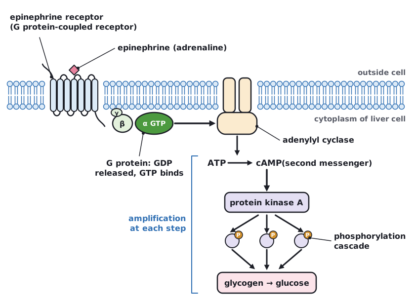 (opens full size in a new tab)
Epinephrine and adrenaline are the same hormone. Both names come from its production by the adrenal gland, above the kidney: an example of naming conventions that persist side by side in international science.
Written and checked against the IB Biology HL specification · Updated October 2026