
Stages in the cardiac cycleSpec B3.2.16
In short
The cardiac cycle is the sequence of events in one heartbeat. The sinoatrial node initiates it: the atria contract (atrial systole), then the ventricles contract (ventricular systole), closing the atrioventricular valves and opening the semilunar valves, then the heart relaxes (diastole). Blood pressure is given as systolic over diastolic, for example 120/80 mm Hg.
The sinoatrial node (the pacemaker) sends out an electrical signal that spreads across the walls of both atria. After a short delay at the atrioventricular node, it spreads through the ventricle walls from the apex (the bottom) upwards. Events in the left side of the heart:
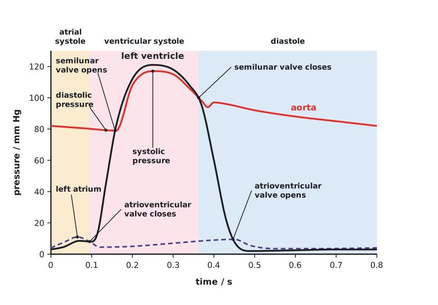
- Atrial systole: the left atrium contracts, raising its pressure slightly above ventricular pressure. The atrioventricular valve is open, so blood is pushed into the left ventricle, topping it up.
- Ventricular systole begins: the ventricle contracts. When ventricular pressure rises above atrial pressure, the atrioventricular valve closes.
- With both valves closed, ventricular pressure rises steeply.
- When ventricular pressure exceeds aortic pressure, the semilunar valve opens and blood is ejected into the aorta.
- Diastole: the ventricle relaxes. When its pressure falls below aortic pressure, the semilunar valve closes, preventing backflow.
- When ventricular pressure falls below atrial pressure, the atrioventricular valve opens and blood flows passively from the left atrium into the ventricle, which fills before the next atrial systole.
 (opens full size in a new tab)
Systolic and diastolic blood pressure
- Systolic pressure is the highest arterial pressure, during ventricular systole.
- Diastolic pressure is the lowest arterial pressure, during diastole; it stays well above zero because the elastic artery walls recoil.
- A typical healthy adult reading is about 120/80 mm Hg (systolic/diastolic). Readings persistently above about 140/90 mm Hg indicate hypertension.
Heart rate from a pressure graph
On a pressure graph, the peaks of ventricular pressure are 0.75 s apart. Calculate the heart rate. (Practice data.)
- One cycle lasts 0.75 s
- Heart rate = 60 ÷ 0.75 = 80
Answer: 80 beats per minute
Valves open and close because of pressure differences on either side of them. Always say which pressure is higher when explaining a valve movement.
Written and checked against the IB Biology HL specification · Updated October 2026