
Gas exchange in leaves

Organisms (Form and function) · Gas exchange · note 4 of 6

Gas exchange in leavesSpec B3.1.7, B3.1.8

In short

A leaf is adapted for gas exchange by stomata, opened and closed by guard cells, which let carbon dioxide in and oxygen out, and by air spaces in the spongy mesophyll that give a large moist surface. The waxy cuticle and epidermis reduce water loss, and veins bring water and remove sugars.

In the light a leaf takes in carbon dioxide for photosynthesis and releases oxygen. The challenge is to let gases in and out without losing too much water.
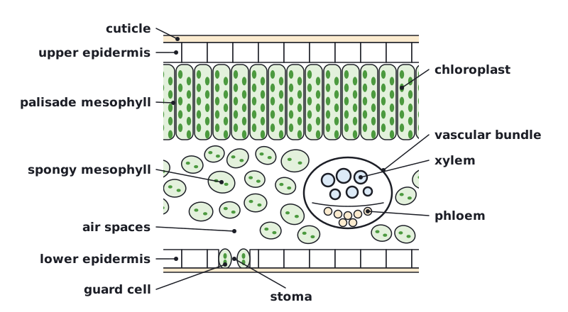
Transverse section of a leaf showing the cuticle, upper epidermis, palisade mesophyll with chloroplasts, spongy mesophyll with air spaces, a vascular bundle with xylem above phloem, the lower epidermis, guard cells and a stoma. (opens full size in a new tab)
Tissues of a dicotyledonous leaf, drawn with cells. A plan diagram shows the same tissues as outlines only.
Adaptations of a leaf for gas exchange
StructureAdaptation
Waxy cuticleWaterproof layer on the epidermis that reduces water loss by evaporation.
EpidermisA single, tightly packed layer of cells that secretes the cuticle and protects the leaf; it is transparent, so light reaches the mesophyll.
Stomatal guard cellsPairs of guard cells open the stoma between them to let CO₂ in and O₂ out, and close it to reduce water loss.
Air spacesConnect the stomata with the mesophyll so gases diffuse quickly through the leaf.
Spongy mesophyllLoosely packed cells with moist walls give a large surface area for gas exchange.
VeinsXylem brings water to replace the water lost; phloem carries away the sugars made in photosynthesis.

Plan diagram of a leaf

A plan diagram shows the distribution of tissues as areas outlined by single lines, with no individual cells drawn. Draw it large, use a sharp pencil, no shading, and label with ruled lines that do not cross.
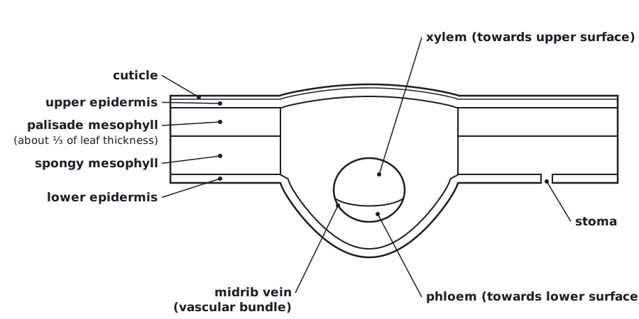
Plan diagram of a transverse section of a dicotyledonous leaf with no cells drawn, labelled cuticle, upper epidermis, palisade mesophyll (about one third of the leaf thickness), spongy mesophyll, lower epidermis with a stoma, and the midrib vein with xylem towards the upper surface and phloem towards the lower surface. (opens full size in a new tab)
Plan diagram of a dicotyledonous leaf: tissues as outlines only. In the vein, xylem is towards the upper surface.
Exam tip:

In the vein, xylem is towards the upper surface and phloem towards the lower surface. Examiners check this in leaf plan diagrams.

Written and checked against the IB Biology HL specification · Updated October 2026

Frequently asked questions

Why do larger organisms need specialised gas exchange surfaces?

Larger organisms need specialised gas exchange surfaces because their surface area-to-volume ratio is smaller and the distance from their centre to the outside is greater. Diffusion across the body surface alone would be far too slow, so they need large, thin, moist, permeable surfaces such as lungs or gills and a transport system.

How are alveoli adapted for gas exchange?

Alveoli are adapted for gas exchange by their huge total surface area, walls one thin layer of cells thick, a moist lining and a dense capillary network around each one. Surfactant lowers surface tension so alveoli do not collapse, and ventilation with continuous blood flow keeps the concentration gradients steep.

What happens to the diaphragm and intercostal muscles when you breathe in?

When you breathe in, the diaphragm contracts and flattens and the external intercostal muscles contract, pulling the ribs up and out. The volume of the thorax increases, so the pressure inside falls below atmospheric pressure and air flows into the lungs.

All 5 questions on Gas exchange