
Tissue fluid and lymphSpec B3.2.11, B3.2.12, B3.2.13
In short
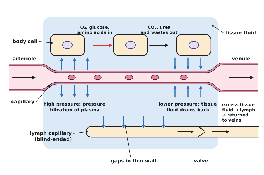
Tissue fluid is formed by pressure filtration of plasma through capillary walls, promoted by high blood pressure from arterioles. It bathes cells, supplying oxygen and glucose and receiving carbon dioxide and wastes. Lower pressure near venules lets most of it drain back into capillaries, and the excess drains into lymph ducts, returning to the blood.
Release and reuptake of tissue fluid
- At the arteriole end of a capillary bed, blood pressure is high.
- Plasma is forced out through gaps in the capillary wall: pressure filtration. This fluid is tissue fluid.
- Large plasma proteins and blood cells stay in the capillary.
- Along the capillary, pressure falls. At the venule end the pressure is lower, so tissue fluid drains back into the capillary (the plasma proteins left in the blood help draw water back in by osmosis).
Exchange between tissue fluid and cells
Tissue fluid bathes every cell. Oxygen, glucose, amino acids and ions diffuse or are transported from the fluid into cells; carbon dioxide, urea and other wastes pass from cells into the fluid and then into the blood.
| Component | Plasma | Tissue fluid |
|---|---|---|
| Water, ions, glucose, amino acids | Present | Present, at similar concentrations |
| Large proteins | High concentration | Very little |
| Oxygen | Higher | Lower (used by cells) |
| Carbon dioxide and wastes | Lower | Higher (released by cells) |
| Red blood cells | Carried in the blood but not part of plasma | Absent |
Drainage into lymph ducts
- Not all tissue fluid returns to the capillaries. The excess drains into blind-ended lymph capillaries, which have thin walls with gaps that let fluid in. The fluid is now lymph.
- Lymph ducts have valves, so lymph flows in one direction only, squeezed along by surrounding muscles.
- Lymph is returned to the blood circulation in veins near the heart (the subclavian veins).
 (opens full size in a new tab)
Written and checked against the IB Biology HL specification · Updated October 2026