
Surface area adaptations and pneumocytesSpec B2.3.7, B2.3.8
In short
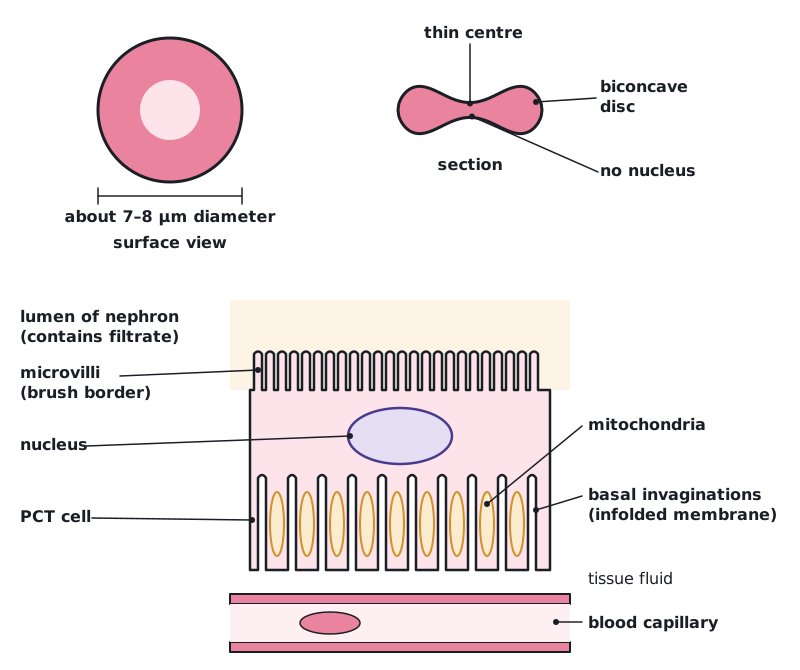
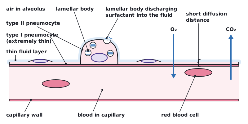
Cells increase their surface area-to-volume ratio by flattening, microvilli and invagination. Erythrocytes are flattened biconcave discs, and proximal convoluted tubule cells have microvilli and an infolded basal membrane. In alveoli, type I pneumocytes are extremely thin to shorten diffusion distances, while type II pneumocytes contain many lamellar bodies that discharge surfactant into the alveolus.
Adaptations that increase SA:V
| Adaptation | Example | How it helps |
|---|---|---|
| Flattening | Erythrocytes (red blood cells) are biconcave discs | A larger surface area for the same volume, and no part of the cytoplasm is far from the surface, so oxygen loads and unloads quickly |
| Microvilli | Proximal convoluted tubule (PCT) cells in the nephron have a brush border of microvilli on the membrane facing the filtrate | Greatly increases the area for reabsorbing substances such as glucose, ions and water from the filtrate |
| Invagination | In PCT cells, the basal membrane facing the tissue fluid and blood capillaries is folded inwards | Increases the area for pumping reabsorbed substances out of the cell; many mitochondria nearby supply ATP |
 (opens full size in a new tab)
Type I and type II pneumocytes
The alveolar epithelium is an example of a tissue with more than one cell type, because different adaptations are needed for the overall function of the tissue.
| Feature | Type I pneumocytes | Type II pneumocytes |
|---|---|---|
| Shape | Extremely thin, flattened cells covering about 95% of the alveolar surface | Rounded (cuboidal) cells; at least as numerous as type I cells but covering only about 5% of the surface |
| Key adaptation | Extreme thinness: the cytoplasm is much less than 1 µm thick | Many secretory vesicles, called lamellar bodies, in the cytoplasm |
| Function | A short distance for diffusion of O₂ and CO₂ between air in the alveolus and blood in the capillaries | Lamellar bodies discharge surfactant into the alveolar lumen; surfactant reduces surface tension in the moist lining, which helps stop the alveoli collapsing |
 (opens full size in a new tab)
Name the vesicles: type II pneumocytes contain lamellar bodies that discharge surfactant. Gas exchange happens mainly across the type I cells.
Written and checked against the IB Biology HL specification · Updated October 2026