
Microscopy skillsSpec A2.2.2
In short
Microscopy skills include making temporary mounts, staining, focusing with the coarse and fine adjustments, measuring with a calibrated eyepiece graticule and taking photographs. Magnification is image size divided by actual size, so actual size equals image size divided by magnification. Always convert both measurements to the same unit, such as micrometres, before dividing.
Making a temporary mount
- Place a thin layer of tissue, such as onion epidermis or cells scraped from the inside of the cheek, on a clean slide in a drop of water or stain.
- Stain to increase contrast: iodine solution for plant cells (stains starch and makes nuclei visible) or methylene blue for animal cells (stains the nucleus).
- Lower a coverslip at an angle with a mounted needle to avoid trapping air bubbles.
- Blot away excess liquid with filter paper.
Focusing
- Start with the low-power objective lens.
- Use the coarse adjustment to bring the specimen roughly into focus.
- Centre the area you want, then rotate to a higher-power objective.
- Use only the fine adjustment at high power, so the lens does not hit and crack the slide.
Measuring with an eyepiece graticule
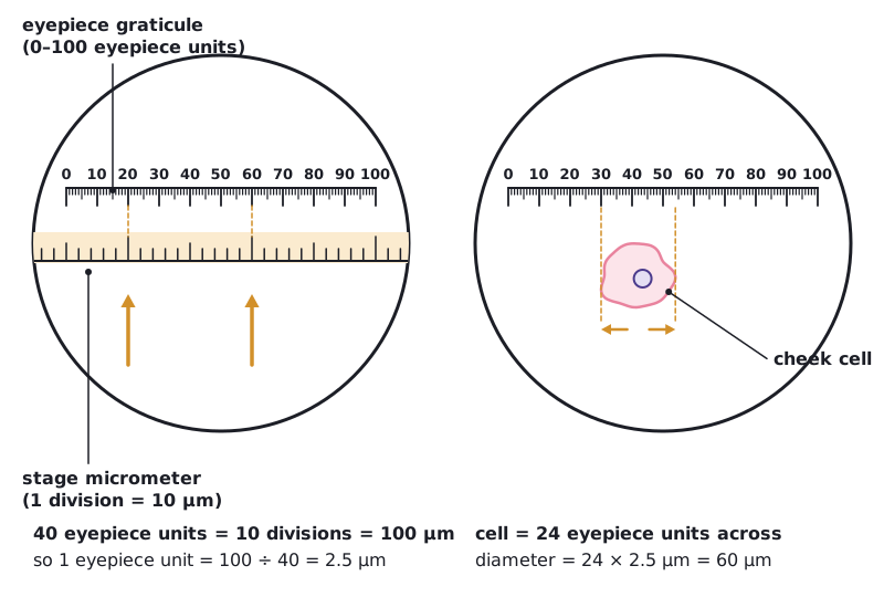
An eyepiece graticule is a scale in the eyepiece marked in arbitrary units. It is calibrated against a stage micrometer, a slide with a ruled scale (often 1 mm divided into 100 divisions of 10 µm). Calibration is repeated for each objective lens, because the length of specimen covered by one eyepiece unit changes with magnification.
Calibrating a graticule
At ×400, 40 eyepiece units line up with 10 divisions of a stage micrometer. Each division is 10 µm. A cheek cell is 24 eyepiece units across. Calculate its actual diameter.
- 10 stage divisions = 10 × 10 µm = 100 µm
- 40 eyepiece units = 100 µm, so 1 eyepiece unit = 100 ÷ 40 = 2.5 µm
- Cell diameter = 24 × 2.5 µm = 60 µm
Answer: 60 µm
 (opens full size in a new tab)
Magnification, actual size and scale bars
Rearranged: actual size = size of image ÷ magnification. Convert to the same units first: 1 mm = 1000 µm and 1 µm = 1000 nm.
Actual size from a micrograph
A Staphylococcus cell is 30 mm across on an electron micrograph with a magnification of ×30 000. Calculate its actual diameter in µm.
- Convert the image size to µm: 30 mm × 1000 = 30 000 µm
- actual size = image size ÷ magnification = 30 000 µm ÷ 30 000
- = 1 µm
Answer: 1 µm
Producing a scale bar
On the same micrograph (×30 000), how long should a scale bar representing 0.5 µm be?
- image size = actual size × magnification
- = 0.5 µm × 30 000 = 15 000 µm
- 15 000 µm ÷ 1000 = 15 mm
Answer: A line 15 mm long, labelled 0.5 µm
To find magnification from a scale bar, measure the bar with a ruler, convert it to the unit written on the bar, then divide the measured length by the value on the bar.
Photographs can be taken through the eyepiece with a camera or phone; add a scale bar so sizes can be measured later. Measurement with an instrument is quantitative observation, so record each reading with its uncertainty.
Written and checked against the IB Biology HL specification · Updated October 2026