
Skeletons, the hip joint and range of motionSpec B3.3.5, B3.3.6, B3.3.7
In short
Skeletons provide anchorage for muscles and act as levers: arthropods have exoskeletons and vertebrates have endoskeletons. At a synovial joint such as the human hip, between the femur and pelvis, cartilage and synovial fluid reduce friction, ligaments hold the bones together and muscles pull on bones through tendons. Range of motion is measured in degrees.
Skeletons as anchorage and levers
- Anchorage: muscles are attached to the skeleton, so when they contract they pull on something firm.
- Levers: rigid parts of the skeleton pivot at joints. A muscle applies force to one point and the bone transmits it to another, changing the size or direction of the force or movement.
- Arthropods (such as insects and crabs) have an exoskeleton on the outside of the body; the muscles are attached to its inner surface.
- Vertebrates have an endoskeleton of bone and cartilage inside the body; muscles are attached to its outer surface by tendons.
Movement at the hip joint
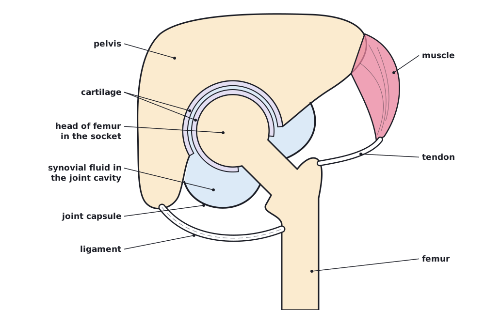
The hip is a ball-and-socket synovial joint: the rounded head of the femur fits into a socket in the pelvis.
| Part | Role |
|---|---|
| Bones (femur and pelvis) | Rigid levers that move relative to each other. |
| Cartilage | Smooth layer covering the bone ends; reduces friction and absorbs shock. |
| Synovial fluid | Lubricates the joint to reduce friction; secreted by the membrane of the joint capsule. |
| Ligaments | Tough bands connecting bone to bone; stabilise the joint and limit movement. |
| Muscles | Contract in antagonistic groups to move the femur. |
| Tendons | Connect muscles to bones and transmit the force of contraction. |
 (opens full size in a new tab)
Range of motion
Range of motion is how far a joint can move, measured as an angle in degrees, in each plane. The hip moves in several dimensions: flexion and extension (forwards and backwards), abduction and adduction (away from and towards the midline) and rotation. A hinge joint such as the knee moves mainly in one plane.
Measure joint angles with a goniometer: place its pivot over the centre of the joint, line one arm up with the fixed bone and the other with the moving bone, and read the angle. Alternatively, photograph or film the movement and measure the angles with image-analysis software. Repeat three times and compare each dimension.
Comparing range of motion
A student measured hip flexion of a classmate three times with a goniometer: 112°, 118° and 115°. Hip abduction was 38°, 42° and 40°. Calculate the mean of each and how many times larger the flexion range is. (Practice data.)
- Mean flexion = (112 + 118 + 115) ÷ 3 = 115°
- Mean abduction = (38 + 42 + 40) ÷ 3 = 40°
- Ratio = 115 ÷ 40 = 2.9
Answer: flexion 115°, abduction 40°; flexion range is about 2.9 times larger
Written and checked against the IB Biology HL specification · Updated October 2026