
Non-specific responses to infectionSpec 6.8
In short
Non-specific responses act against any pathogen in the same way. Inflammation increases blood flow and brings phagocytes to damaged tissue, lysozyme digests bacterial cell walls, interferon stops viruses replicating in neighbouring cells, and phagocytosis engulfs and digests pathogens in phagocytes.
Inflammation
When tissue is damaged or infected, mast cells and damaged cells release histamine and other signalling molecules (cytokines).
- Histamine causes vasodilation of arterioles, increasing blood flow to the area, so the tissue becomes red and warm.
- Capillary walls become more permeable, so more fluid leaves the blood as tissue fluid, causing swelling, which may press on nerve endings and cause pain.
- Chemical signals attract phagocytes (neutrophils and monocytes that become macrophages), which leave the capillaries and engulf pathogens.
Lysozyme
Lysozyme is an enzyme found in tears, saliva, mucus and in phagocytes. It hydrolyses the peptidoglycan in the cell walls of bacteria. The weakened wall can no longer resist osmotic pressure, so water enters by osmosis and the bacterium bursts (lysis). Lysozyme does not act on viruses.
Interferon
Interferons are cytokines (signalling proteins) released by cells infected with a virus. They diffuse to neighbouring uninfected cells and bind to receptors on the cell-surface membrane. This switches on genes that make enzymes that break down viral mRNA and inhibit protein synthesis. Viral proteins cannot be made, so the virus cannot replicate in these cells and its spread is slowed.
Phagocytosis
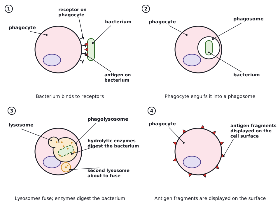
- A phagocyte (neutrophil or macrophage) recognises a pathogen, often because it is coated with antibodies or has foreign surface molecules, and the pathogen binds to receptors on the phagocyte.
- The phagocyte engulfs the pathogen by endocytosis, enclosing it in a vesicle called a phagosome.
- Lysosomes fuse with the phagosome and release lysozyme and other hydrolytic enzymes, which digest the pathogen.
- Soluble products are absorbed into the cytoplasm. A macrophage then displays pieces of the pathogen's antigens on its surface (antigen presentation) to start the specific response.
'Non-specific' means the response is the same whatever the pathogen. Do not say it is 'not immune': it is the first part of the innate immune response.
 (opens full size in a new tab)
Written and checked against the Edexcel International A Level Biology (YBI11) specification · Updated October 2026