
Resting potential and action potentialsSpec 8.4
In short
An action potential is a brief reversal of the potential difference across an axon membrane. Sodium channels open, so sodium ions enter and depolarise it; they then close and potassium channels open, so potassium ions leave and repolarise it. Local currents trigger the next region, so the impulse travels along the axon.
At rest, the inside of an axon is negative relative to the outside. This resting potential is about –70 mV. It is set up by the sodium–potassium pump, which uses ATP to move 3 Na⁺ out of the axon for every 2 K⁺ moved in, and by potassium ion channels that leak K⁺ out far more easily than Na⁺ can leak in. The membrane is said to be polarised.
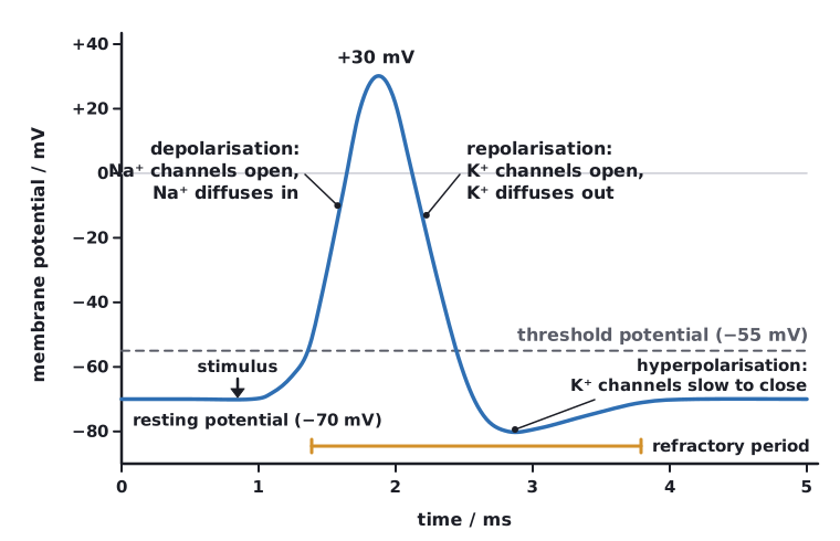
A stimulus that is strong enough opens some voltage-gated sodium ion channels. When the membrane potential reaches the threshold (about –55 mV), many more open. This is an all-or-nothing response: below threshold nothing happens, and above it every action potential has the same size.
- Depolarisation: voltage-gated Na⁺ channels open, so Na⁺ diffuses in down its electrochemical gradient. The inside becomes positive, reaching about +40 mV.
- At the peak the Na⁺ channels close (they are inactivated) and voltage-gated K⁺ channels open.
- Repolarisation: K⁺ diffuses out of the axon, so the inside becomes negative again.
- Hyperpolarisation: the K⁺ channels are slow to close, so the potential briefly falls below –70 mV. The K⁺ channels then close and the sodium–potassium pump and K⁺ leak channels restore the resting potential.
 (opens full size in a new tab)
During and just after an action potential the Na⁺ channels are inactivated, so the region cannot respond to another stimulus. This refractory period means impulses travel in one direction only, do not overlap, and are separate events. The frequency of impulses, not their size, carries information about stimulus strength.
An action potential travels along the axon by local circuits. Na⁺ entering one region diffuses sideways inside the axon, depolarising the next region to threshold, so its Na⁺ channels open. The region behind is refractory, so the impulse moves forward.
Use the terms ‘voltage-gated’, ‘down an electrochemical gradient’ and ‘permeability’. State which ion moves, in which direction, through which channel, and what happens to the potential.
Written and checked against the Edexcel International A Level Biology (YBI11) specification · Updated October 2026