
Imaging the brain: CT, MRI, fMRI and PETSpec 8.15
In short
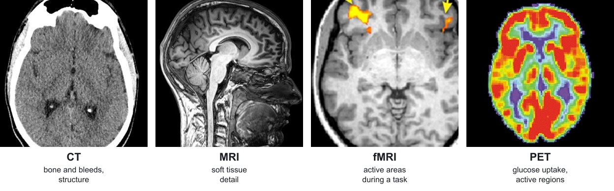
CT uses X-rays to build images of brain structure, and MRI uses magnetic fields and radio waves to give detailed soft-tissue images. fMRI detects changes in blood oxygen to map active areas, and PET uses a radioactive tracer to show where the brain uses glucose. They diagnose disease and investigate brain function.
| Technique | How it works | What it shows and uses | Advantages and limitations |
|---|---|---|---|
| CT (computed tomography) | An X-ray source rotates around the head. Detectors measure how much is absorbed by tissues; a computer builds cross-sectional slices. | Brain structure: bone, tumours, bleeding in stroke or head injury | Quick and widely available. Uses ionising radiation; poorer soft-tissue contrast than MRI; shows structure, not activity. |
| MRI (magnetic resonance imaging) | A strong magnetic field aligns hydrogen nuclei in water. A pulse of radio waves knocks them out of line; they emit signals as they return, which differ between tissues. | Detailed images of soft tissue: tumours, damage to brain tissue, multiple sclerosis, structural abnormalities | No ionising radiation and high resolution. Slow, noisy, expensive; cannot be used with some metal implants or pacemakers. |
| fMRI (functional MRI) | MRI that detects changes in blood oxygen. Active neurones use more oxygen, so blood flow to them increases, which changes the signal. | Which parts of the brain are active during a task, such as speaking or moving a hand | Shows function as well as structure and uses no radiation. Indirect measure of activity; expensive; the person must keep still. |
| PET (positron emission tomography) | A radioactive tracer, such as a glucose analogue labelled with a positron emitter, is injected. Active cells take up more of it. Detectors record the gamma rays produced. | Metabolic activity, such as areas that are abnormally active or inactive in epilepsy, dementia or tumours | Shows chemical activity. Uses radioactivity; costly; the tracer has a short half-life, so it must be made close to the scanner. |
 (opens full size in a new tab)
Exam tip:
Choose the scan by what the question asks: structure (CT or MRI) or function (fMRI or PET). Then give a reason and a limitation, because ‘evaluate’ questions need both.
Written and checked against the Edexcel International A Level Biology (YBI11) specification · Updated October 2026