
Rods, rhodopsin and the optic neuroneSpec 8.8
In short
Rod cells in the retina contain rhodopsin, made of opsin and retinal. In the dark, cation channels are open, Na⁺ enters and the rod is depolarised. Light splits rhodopsin, the channels close, the rod hyperpolarises and releases less transmitter, so a bipolar neurone triggers action potentials in the optic neurone.
Rod cells in the retina detect light, particularly in dim conditions. Their outer segment contains the pigment rhodopsin, made of a protein, opsin, bound to retinal, a derivative of vitamin A.
In the retina, the rods synapse with bipolar neurones, which in turn synapse with the optic neurones whose axons form the optic nerve. The key point is that light causes hyperpolarisation, not depolarisation, of the rod cell.
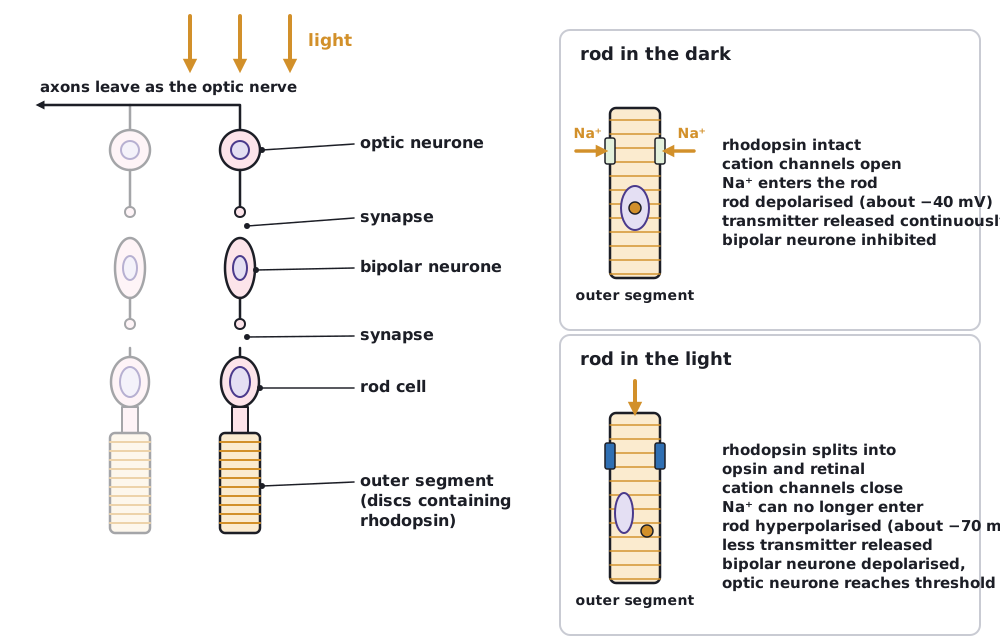 (opens full size in a new tab)
- In the dark: cation channels in the rod membrane are open, so Na⁺ continuously enters. The rod is partly depolarised (about –40 mV) and releases an inhibitory transmitter (glutamate) onto the bipolar neurone, so the bipolar neurone is hyperpolarised and does not pass a signal on.
- In the light: absorbing light changes retinal from its cis form to its trans form, so rhodopsin breaks down into opsin and retinal (bleaching). Opsin activates a cascade of reactions in the membrane (a fall in cyclic GMP, which normally keeps the channels open), so the cation channels close.
- Na⁺ can no longer enter, but the sodium–potassium pump keeps removing it and K⁺ continues to leave, so the rod becomes more negative: hyperpolarisation (about –70 mV).
- The hyperpolarised rod releases less inhibitory transmitter, so the bipolar neurone is depolarised and releases transmitter onto an optic neurone.
- The optic neurone reaches threshold and action potentials pass along the optic nerve to the visual cortex of the brain.
Rods are very sensitive because one photon can close many channels. Several rods synapse with one bipolar neurone, which makes rods sensitive in dim light but gives low visual acuity, and they do not distinguish colours.
The rod cell itself does not fire action potentials. The action potentials are formed in the optic neurone, after the bipolar neurone is depolarised.
Written and checked against the Edexcel International A Level Biology (YBI11) specification · Updated October 2026