
How the pupil dilates and constricts

Coordination, Response and Gene Technology · Impulses, synapses and receptors · note 4 of 7

Spec 8.6
A Level only (what this means)A Level only: only in the full A Level, not the AS Level. AS Level students can skip it. What the labels mean
Download all Impulses, synapses and receptors notes (PDF)Download all notes (PDF)8 pages

How the pupil dilates and constrictsSpec 8.6

In short

The pupil changes size because circular and radial muscles in the iris work antagonistically. In bright light, circular muscles contract and radial muscles relax, so the pupil constricts and protects the retina. In dim light, radial muscles contract and circular muscles relax, so the pupil dilates and lets in more light.

The iris contains two sets of muscle that act as effectors, controlling the size of the pupil and so how much light reaches the retina. They are antagonistic muscles: when one contracts the other relaxes.

Pupil responses
Bright lightDim light
Circular musclesContractRelax
Radial musclesRelaxContract
PupilConstricts (gets smaller)Dilates (gets larger)
PurposeProtects the retina (the light receptors) from damage by intense lightLets more light reach the retina so rods can detect it
ControlParasympathetic neurones release acetylcholine onto the circular musclesSympathetic neurones release noradrenaline onto the radial muscles

The pupil constriction in bright light is a reflex. Light receptors in the retina send impulses along the optic nerve to the brain, and motor neurones carry impulses to the muscles of the iris. This is rapid and automatic, so it protects the retina.
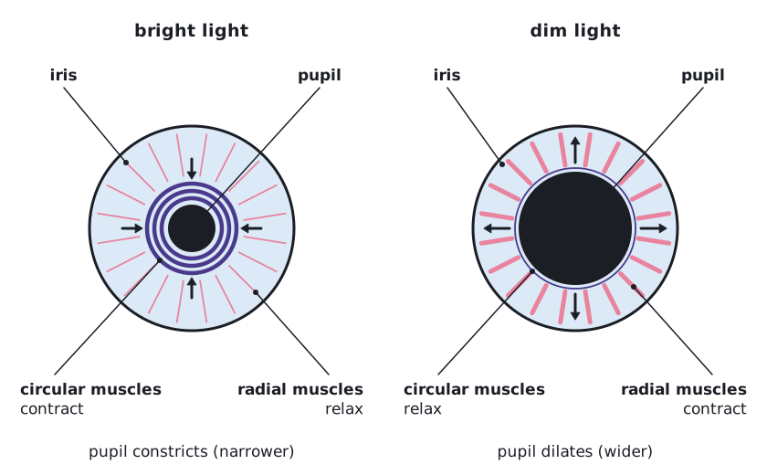
Front view of the eye in bright and dim light showing iris, pupil, circular muscles and radial muscles and the size of the pupil in each. (opens full size in a new tab)
In bright light the circular muscles contract and the pupil constricts; in dim light the radial muscles contract and it dilates.
Common mistake:

Do not say that the iris ‘opens and closes’. The pupil is the hole; the muscles in the iris change its size.

Written and checked against the Edexcel International A Level Biology (YBI11) specification · Updated October 2026

Frequently asked questions

How does an action potential work?

An action potential is a brief reversal of the potential across an axon membrane. Voltage-gated sodium channels open at threshold, so Na⁺ enters and the membrane depolarises. The sodium channels then close and potassium channels open, so K⁺ leaves and the membrane repolarises. Local circuits depolarise the next region, so the impulse travels.

Why do myelinated axons conduct impulses faster?

Myelin insulates the axon so ions cross the membrane only at the nodes of Ranvier. The action potential jumps from node to node by local circuits, which is called saltatory conduction. Less of the membrane has to depolarise, so conduction reaches about 100 m s⁻¹, much faster than the 0.5 to 2 m s⁻¹ in an unmyelinated axon.

How does acetylcholine transmit a signal across a synapse?

An impulse opens calcium channels in the presynaptic knob, and calcium ions make vesicles release acetylcholine by exocytosis. Acetylcholine diffuses across the cleft and binds to receptors on the postsynaptic membrane, opening sodium channels. Sodium ions enter and depolarise it. Acetylcholinesterase then breaks the acetylcholine down.

All 5 questions on Impulses, synapses and receptors