
Structure of a skeletal muscle fibreSpec 7.10(i)
In short
A mammalian skeletal muscle fibre is a long, multinucleate cell with a cell surface membrane called the sarcolemma and cytoplasm called sarcoplasm. It is packed with myofibrils, each made of repeating sarcomeres containing thick myosin and thin actin filaments, and it has many mitochondria, sarcoplasmic reticulum and T-tubules.
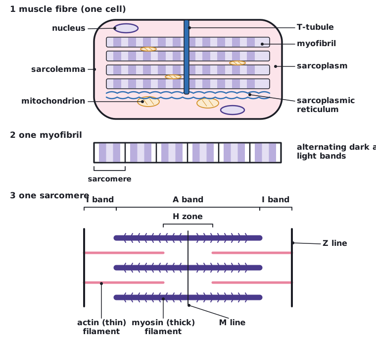
A skeletal muscle fibre is a single, long, cylindrical cell with many nuclei. Its cell surface membrane is the sarcolemma and its cytoplasm is the sarcoplasm. The sarcolemma folds inwards to form T-tubules (transverse tubules), which carry action potentials deep into the fibre.
- Myofibrils: long, thin, cylindrical bundles of protein filaments that run along the length of the fibre and cause contraction.
- Sarcoplasmic reticulum: a network of membranes around each myofibril that stores calcium ions (Ca²⁺).
- Many mitochondria: lying between the myofibrils, to supply the ATP needed for contraction.
- Many nuclei, lying just under the sarcolemma.
The sarcomere
Each myofibril is divided along its length into repeating units called sarcomeres, from one Z line to the next. Two kinds of filament lie in the sarcomere: thick filaments of myosin and thin filaments of actin (with the proteins troponin and tropomyosin). Their arrangement gives the striped (striated) appearance under the microscope.
| Region | Appearance | What is present |
|---|---|---|
| A band | Dark | The full length of the myosin filaments (including where actin overlaps them) |
| I band | Light | Actin filaments only (no myosin) |
| H zone | Lighter region in the middle of the A band | Myosin only (no actin overlap) |
| Z line | Thin dark line | Boundary of the sarcomere; actin is attached here |
| M line | Centre of the sarcomere | Myosin filaments held together |
 (opens full size in a new tab)
In a contracted muscle the A band stays the same length, but the I band and H zone get shorter. Examiners often test this.
Written and checked against the Edexcel International A Level Biology (YBI11) specification · Updated October 2026