
Arteries, veins and capillariesSpec 1.7
In short
Arteries have thick, elastic, muscular walls to withstand high pressure and recoil to smooth the pulse. Veins have thin walls, wide lumens and valves to return blood at low pressure. Capillaries have walls one cell thick and a narrow lumen, giving a short diffusion distance and slow flow for exchange with cells.
| Feature | Artery | Vein | Capillary |
|---|---|---|---|
| Direction of blood flow | Away from the heart | Towards the heart | Between arteries and veins (through tissues) |
| Blood pressure | High | Low | Falls along the capillary |
| Wall | Thick, with collagen, smooth muscle and elastic fibres | Thin, with little muscle or elastic tissue | One layer of flattened endothelial cells |
| Lumen | Narrow | Wide | Very narrow (about the width of a red blood cell) |
| Valves | None (except at the heart) | Present | None |
| Main function | Carry blood away from the heart at high pressure | Return blood to the heart | Exchange of substances between blood and cells |
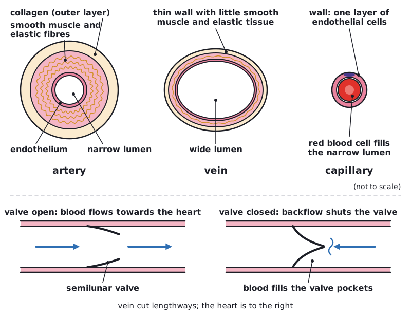 (opens full size in a new tab)
Arteries
Blood leaves the heart in a high-pressure surge. The wall of an artery contains collagen fibres, which give strength and prevent bursting, and elastic fibres, which stretch as the surge passes and then recoil. This elastic recoil evens out the pressure pulse so blood flows smoothly. Smooth muscle in the wall can contract to narrow the lumen. The inner lining (endothelium) is smooth, which reduces friction.
Veins
By the time blood reaches the veins its pressure is low, so the walls can be thin, with little muscle or elastic tissue. The wide lumen offers little resistance to flow. Valves (semilunar valves) stop blood flowing backwards. Blood is helped towards the heart by contractions of the surrounding skeletal muscles squeezing the veins, and by pressure changes in the chest during breathing.
Capillaries
Capillaries form dense networks close to almost every cell. Their wall is a single layer of flattened endothelial cells, so the diffusion distance is very short. Gaps between the cells allow small molecules and fluid to pass. The lumen is so narrow that red blood cells are squeezed through in single file, which brings them close to the wall. Together, the capillaries have a very large total cross-sectional area, so blood flows slowly. This gives time for exchange of oxygen, carbon dioxide, glucose and wastes.
Explain each feature with 'so': thick elastic wall so it withstands high pressure and recoils; one cell thick so a short diffusion distance. A list of features without the reason will not get the mark.
Written and checked against the Edexcel International A Level Biology (YBI11) specification · Updated October 2026