
Cardiac output and the control of heart rate and ventilationSpec 7.13
In short
Cardiac output is the volume of blood pumped by a ventricle per minute, calculated as stroke volume multiplied by heart rate. During exercise, chemoreceptors detect rising carbon dioxide and falling pH, and the cardiovascular control centre and ventilation centre in the medulla oblongata increase heart rate and ventilation to deliver oxygen and remove carbon dioxide rapidly.
Stroke volume is the volume of blood pumped out of a ventricle in one beat (cm³). Heart rate is in beats per minute. Cardiac output is in cm³ min⁻¹ (or dm³ min⁻¹).
Cardiac output
At rest, a person's stroke volume is 75 cm³ and heart rate 70 beats per minute. During exercise, stroke volume is 120 cm³ and heart rate 150 beats per minute. Calculate the change in cardiac output.
- Resting cardiac output = 75 × 70 = 5250 cm³ min⁻¹
- Exercise cardiac output = 120 × 150 = 18 000 cm³ min⁻¹
- Change = 18 000 − 5250
Answer: 12 750 cm³ min⁻¹ (12.75 dm³ min⁻¹) increase. An increase of 243% (12 750 ÷ 5250 × 100).
Write the unit with every answer and check the units match (cm³ with cm³ min⁻¹). Percentage change = change ÷ original × 100.
Controlling heart rate
The cardiovascular control centre in the medulla oblongata of the brain receives information from receptors and signals to the SAN along two nerves:
- Chemoreceptors in the carotid arteries and aorta, and in the medulla, detect a fall in blood pH (caused by a rise in CO₂ concentration, which forms carbonic acid). Baroreceptors in the aorta and carotid arteries detect blood pressure.
- A rise in CO₂ (low pH) in exercise: impulses along sympathetic nerves (the accelerator pathway) to the SAN increase heart rate and stroke volume.
- A rise in blood pressure, or low CO₂: impulses along the vagus (parasympathetic) nerve to the SAN decrease heart rate.
- During exercise, stretch receptors in the muscles and joints also detect movement and send impulses to the cardiovascular control centre, so heart rate rises as soon as exercise starts. Adrenaline released into the blood increases heart rate as well.
Controlling ventilation
The ventilation centre (breathing centre) in the medulla oblongata controls the rate and depth of breathing. During exercise, more CO₂ is made by respiration, and chemoreceptors detect the fall in pH. The ventilation centre sends more frequent impulses along nerves to the diaphragm and the external intercostal muscles, so breathing becomes faster and deeper. Stretch receptors in the lungs send impulses back to the centre to stop over-inflation.
The combined increase in ventilation and cardiac output delivers oxygen to the muscles quickly and removes CO₂ rapidly from them, keeping the blood CO₂ concentration and pH within narrow limits. This is a negative feedback system.
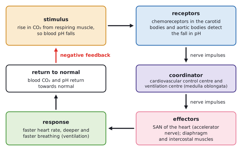 (opens full size in a new tab)
Written and checked against the Edexcel International A Level Biology (YBI11) specification · Updated October 2026