
Levels of protein structure and the bonds involvedSpec 2.6
In short
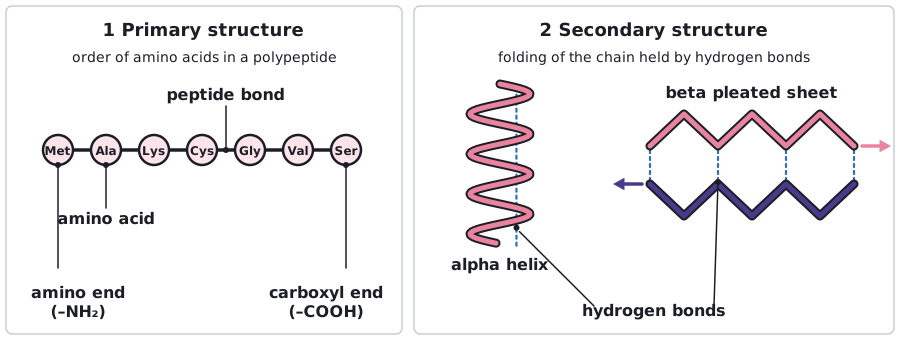
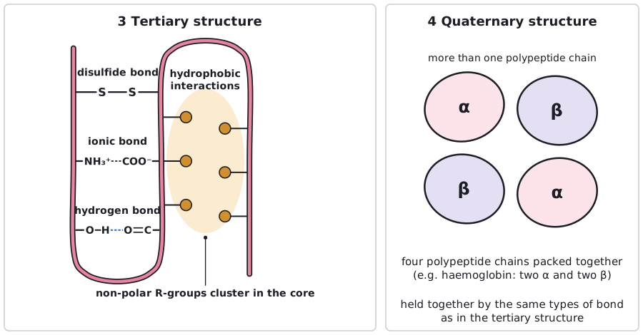
The primary structure of a protein is its sequence of amino acids. It determines how the chain coils or folds into the secondary structure (alpha helix or beta pleated sheet) and then the tertiary structure, held by hydrogen, ionic and disulfide bonds. Several polypeptides together form a protein's quaternary structure.
| Level | Description | Bonds involved |
|---|---|---|
| Primary | The sequence of amino acids in the polypeptide chain. | Peptide bonds (covalent). |
| Secondary | Regular coiling or folding of the chain: the alpha helix and the beta pleated sheet. | Hydrogen bonds between the C=O and N–H groups of the polypeptide backbone, between amino acids near each other. |
| Tertiary | Further folding of the whole chain into a specific three-dimensional shape. | Hydrogen bonds, ionic bonds, disulfide bonds (covalent, between sulfur atoms of cysteine residues), and hydrophobic interactions between non-polar R groups. |
| Quaternary | Two or more polypeptide chains (and sometimes a non-protein group) together in one protein. | The same types of bond and interaction between the chains. |
The primary structure decides everything else. The sequence of R groups determines where hydrogen bonds, ionic bonds and disulfide bonds can form, so it determines how the chain folds, and therefore the final shape and properties of the protein. Changing even one amino acid can change the shape, and so the function.
The bonds in the tertiary structure
- Hydrogen bonds: weak, but numerous. They form between polar R groups. They are broken by changes in temperature and pH.
- Ionic bonds: between positively and negatively charged R groups (for example –NH₃⁺ and –COO⁻). Stronger than hydrogen bonds but broken by changes in pH.
- Disulfide bonds: covalent bonds between the sulfur atoms of two cysteine R groups. Strong, and not easily broken.
- Hydrophobic interactions: non-polar R groups cluster in the centre of a globular protein, away from water.
Do not say the primary structure is 'held together by hydrogen bonds'. Peptide bonds hold the primary structure; hydrogen bonds hold the secondary structure and help hold the tertiary structure.
 (opens full size in a new tab)
 (opens full size in a new tab)
Written and checked against the Edexcel International A Level Biology (YBI11) specification · Updated October 2026