
Structure of the mammalian kidneySpec 7.18
In short
The mammalian kidney has an outer cortex, an inner medulla and a pelvis leading to the ureter. Each kidney contains many nephrons: a Bowman's capsule containing a glomerulus, a proximal convoluted tubule, a loop of Henle, a distal convoluted tubule and a collecting duct. Blood arrives in the renal artery and leaves in the renal vein.
Gross structure
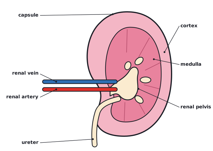
- Renal artery brings oxygenated blood with urea; renal vein carries blood away.
- Fibrous capsule around the outside.
- Cortex: outer region, containing the glomeruli, Bowman's capsules and convoluted tubules.
- Medulla: inner region, containing the loops of Henle and collecting ducts.
- Renal pelvis: the funnel-shaped cavity that collects urine and drains into the ureter, which leads to the bladder.
Microscopic structure: the nephron
| Part | Position | Structure and role |
|---|---|---|
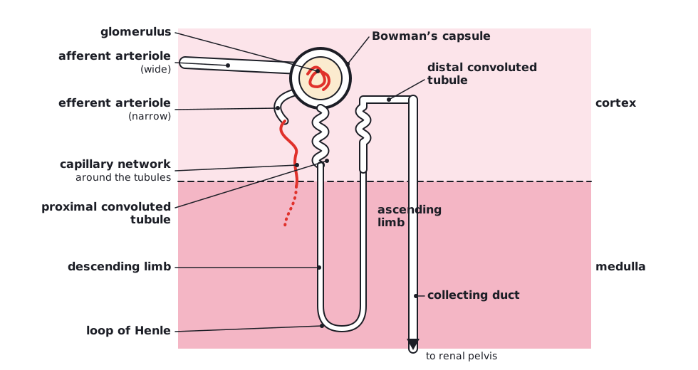
| Glomerulus | Cortex (inside Bowman's capsule) | Knot of capillaries; the afferent arteriole is wider than the efferent arteriole, which creates high pressure |
| Bowman's capsule | Cortex | Cup-shaped; the inner layer has podocytes; collects the filtrate |
| Proximal convoluted tubule | Cortex | Cuboidal epithelium with microvilli and many mitochondria; reabsorbs most of the filtrate |
| Loop of Henle | Medulla (descends and ascends) | Creates a high solute concentration (low water potential) in the medulla |
| Distal convoluted tubule | Cortex | Adjusts the ion content of the filtrate |
| Collecting duct | Cortex to medulla to pelvis | Collects urine from many nephrons; water reabsorbed under the control of ADH |
Blood flows from the renal artery to the afferent arteriole, glomerulus and efferent arteriole, then through a capillary network around the tubules (where substances are reabsorbed), and leaves in the renal vein.
 (opens full size in a new tab)
 (opens full size in a new tab)
Written and checked against the Edexcel International A Level Biology (YBI11) specification · Updated October 2026