
Cardiac muscle and the coordination of the heartbeat

Respiration, Muscles and the Internal Environment · The heart, ventilation and exercise · note 1 of 5

Spec 7.12(i), 7.12(ii)
A Level only (what this means)A Level only: only in the full A Level, not the AS Level. AS Level students can skip it. What the labels mean
Download all The heart, ventilation and exercise notes (PDF)Download all notes (PDF)7 pages

Cardiac muscle and the coordination of the heartbeatSpec 7.12(i), 7.12(ii)

In short

Cardiac muscle is myogenic: it contracts and relaxes rhythmically without nervous stimulation. The sinoatrial node sets the pace and sends a wave of excitation across the atria; the atrioventricular node delays it, then the bundle of His and Purkyne fibres carry it to the ventricles, which contract from the apex upwards.

Cardiac muscle is myogenic: it can initiate its own contraction without a nerve impulse, so the heart beats even if its nerves are cut. Nerves only change the rate.

  1. The sinoatrial node (SAN), a patch of cardiac muscle cells in the wall of the right atrium, is the pacemaker. It generates a wave of depolarisation (excitation) at regular intervals.
  2. The wave spreads across the walls of both atria, which contract together (atrial systole).
  3. A layer of non-conducting fibrous tissue between the atria and the ventricles stops the wave passing directly to the ventricles.
  4. The wave reaches the atrioventricular node (AVN). The AVN delays it for about 0.1 s, giving the atria time to empty and the ventricles time to fill.
  5. The wave then passes down the bundle of His, which runs through the septum to the apex (bottom) of the heart.
  6. The bundle divides into Purkyne fibres, which carry the wave up the walls of both ventricles. The ventricles contract from the apex upwards (ventricular systole), forcing blood out through the arteries.
Exam tip:

Explain the delay at the AVN: it makes sure the atria have finished contracting before the ventricles contract, so the ventricles fill fully.
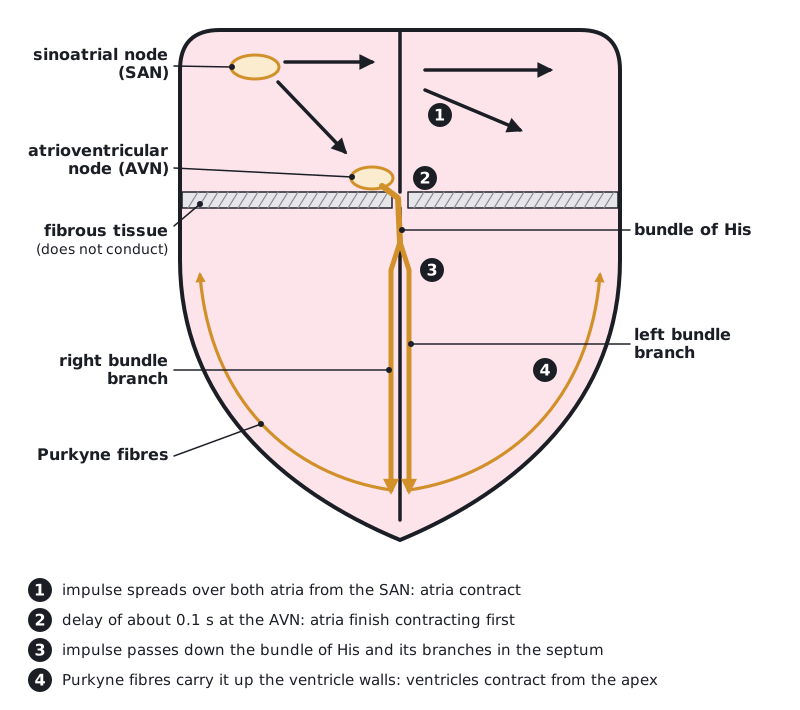
Longitudinal section of the heart showing the SAN in the right atrial wall, fibrous non-conducting tissue between atria and ventricles, the AVN, bundle of His, left and right bundle branches and Purkyne fibres, with numbered stages 1 to 4 and a 0.1 s delay at the AVN. (opens full size in a new tab)
The wave of excitation: SAN, then a 0.1 s delay at the AVN, then the bundle of His, branches and Purkyne fibres.

Written and checked against the Edexcel International A Level Biology (YBI11) specification · Updated October 2026

Frequently asked questions

How does the heart beat without being told by the brain?

Cardiac muscle is myogenic, so it generates its own excitation. The sinoatrial node in the right atrium sets the pace and sends a wave across the atria. The atrioventricular node delays it, then the bundle of His and Purkyne fibres spread it through the ventricles. Nerves only change the rate.

How do you read an ECG?

The P wave is atrial depolarisation, the QRS complex is ventricular depolarisation and the T wave is ventricular repolarisation. The time between two R peaks gives the heart rate: 60 divided by that time in seconds. Irregular spacing or missing P waves can show abnormal rhythms such as atrial fibrillation.

What controls heart rate and breathing rate during exercise?

The medulla oblongata does. Chemoreceptors detect a rise in carbon dioxide and a fall in pH in the blood. The cardiovascular control centre increases heart rate through the sympathetic nerve to the sinoatrial node, and the ventilation centre speeds up and deepens breathing, to deliver oxygen and remove carbon dioxide.

All 5 questions on The heart, ventilation and exercise