
Separating DNA fragments by gel electrophoresisSpec 6.18
In short
Gel electrophoresis separates DNA fragments by length. DNA is negatively charged because of its phosphate groups, so in an electric field it moves through an agarose gel towards the positive electrode. Shorter fragments move faster and further, and fragments are made visible with a fluorescent dye or probe.
- DNA is cut into fragments with restriction enzymes (or amplified by PCR).
- A gel made of agarose (a porous polysaccharide) is covered with a buffer solution that conducts electricity. Samples, mixed with loading dye to make them dense and visible, are put into wells at the negative end. A mixture of fragments of known lengths (a DNA ladder) is put in one well.
- An electric current is applied. DNA is negatively charged because of the phosphate groups, so it moves through the gel towards the positive electrode (anode).
- The gel acts as a mesh. Shorter fragments move through it more easily, so they travel further in a given time, and longer fragments move more slowly. Fragments of the same length form a band.
- The DNA is made visible by staining with a fluorescent dye that binds to DNA and is viewed in UV light, or by DNA probes that carry a fluorescent or radioactive label.
The length of a fragment is estimated by comparing how far its band has moved with the bands of the DNA ladder. A graph of log₁₀ fragment length against distance moved is roughly a straight line.
The two key ideas are charge (negative phosphate groups, so moves to the anode) and size (smaller fragments move further through the gel). Fragments are separated by length, not by mass of the whole sample.
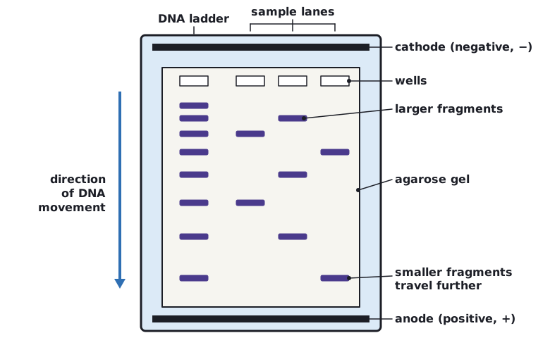 (opens full size in a new tab)
Written and checked against the Edexcel International A Level Biology (YBI11) specification · Updated October 2026