
Core Practical 5: observing, drawing and measuring cellsSpec 3.8
In short
In Core Practical 5, animal cells such as stained cheek cells are viewed with a light microscope and drawn with clear, labelled outlines. An eyepiece graticule is calibrated against a stage micrometer at each objective lens, so that the actual size of a cell can be calculated from the number of graticule divisions it covers.
Core practical 5: use a light microscope to make observations and labelled drawings of suitable animal cells, and use a graticule to make measurements and understand scale.
Preparing and viewing the slide
- Wash hands. Use a clean cotton bud (or a sterile spatula) to gently scrape the inside of your own cheek.
- Smear the cells onto a clean microscope slide in a drop of water or stain (for example methylene blue). Dispose of the swab in disinfectant.
- Lower a coverslip at an angle with a mounted needle, to avoid trapping air bubbles. Blot excess stain with filter paper.
- Start with the lowest-power objective lens, focus with the coarse focus and then the fine focus, and increase the magnification as needed.
- Alternatively, use a prepared slide of an animal tissue, such as a section of liver or of squamous epithelium.
Making a labelled drawing
- Use a sharp pencil, a clear continuous line, no shading and no colouring.
- Draw only what you see. Make the drawing large (fill at least half of the space) and draw a few cells, not the whole field of view.
- Label with ruled lines that do not cross and touch the structure, and give the title, the magnification used and a scale bar.
Measuring with a graticule
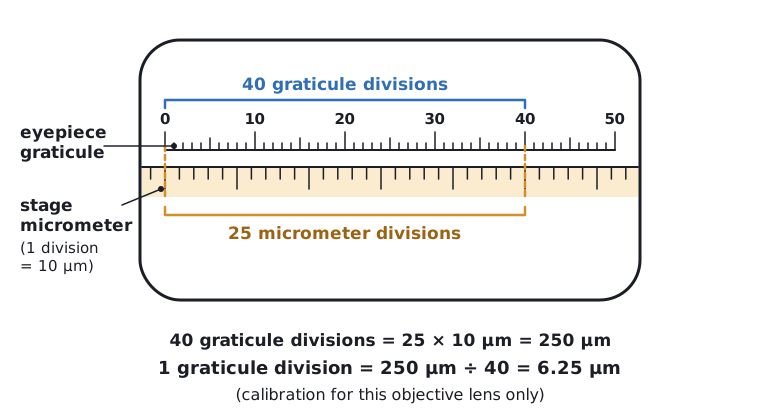
An eyepiece graticule is a transparent scale with arbitrary divisions placed in the eyepiece. Its units have no fixed size, so it must be calibrated for each objective lens against a stage micrometer, a slide with a scale of known size (usually divisions of 10 µm).
- Place the stage micrometer on the stage and focus on its scale.
- Line up the zero of the graticule with the zero of the micrometer.
- Find a point further along where two divisions line up again. Count the graticule divisions and the micrometer divisions between these points.
- Calculate the size of one graticule division in µm.
- Replace the micrometer with the specimen and measure the cell in graticule divisions. Multiply by the calibration value. Repeat for several cells and calculate a mean.
Calibrating a graticule
At ×400, 40 graticule divisions are the same length as 25 divisions of a stage micrometer. Each micrometer division is 10 µm. A cell measures 12 graticule divisions. Calculate the actual length of the cell.
- Length of 25 micrometer divisions = 25 × 10 = 250 µm
- One graticule division = 250 ÷ 40 = 6.25 µm
- Cell length = 12 × 6.25
Answer: 75 µm
Recalibrate whenever the objective lens is changed. Measuring several cells and finding the mean makes the result more reliable. Work in µm and convert to mm by dividing by 1000.
Risks and controls: your own cheek cells should not be shared and must be disposed of in disinfectant, to prevent the spread of infection. Stains such as methylene blue can stain skin and clothes. Take care with glass slides and coverslips, which can break. Wear eye protection if the stain is an irritant.
 (opens full size in a new tab)
Quick check
Define resolution.
Show answer
The smallest distance between two points at which they can still be seen as separate.
Why do electron microscopes have a higher resolution than light microscopes?
Show answer
Electrons have a much shorter wavelength than light.
Why are specimens stained?
Show answer
To increase contrast so that structures are visible and can be told apart.
What is the formula for magnification?
Show answer
Magnification = size of image ÷ size of real object.
Why must an eyepiece graticule be calibrated?
Show answer
Its divisions have no fixed size; they depend on the objective lens, so they must be compared with a stage micrometer.
Written and checked against the Edexcel International A Level Biology (YBI11) specification · Updated October 2026