
Magnification and resolutionSpec 3.7
In short
Magnification is how many times larger an image is than the object. Resolution is the smallest distance between two points that can still be seen as separate. Light microscopes resolve about 200 nm because of the wavelength of light, whereas electron microscopes use electrons with a much shorter wavelength, so they resolve far smaller structures.
- Magnification
- How many times larger the image is than the real object.
- Resolution
- The smallest distance between two points at which they can still be seen as two separate points.
Magnification can be increased as much as you like, but the image only shows more detail if the resolution is good enough. Resolution is limited by the wavelength of the radiation used: two points closer together than about half the wavelength merge into one.
| Feature | Light microscope | Transmission electron microscope (TEM) | Scanning electron microscope (SEM) |
|---|---|---|---|
| Radiation | Light (wavelength about 400 to 700 nm), focused by glass lenses | Beam of electrons (very short wavelength), focused by electromagnets, in a vacuum | Beam of electrons scanned over the surface, in a vacuum |
| Maximum useful magnification | About ×1500 | Over ×1 000 000 | Up to about ×500 000 |
| Resolution | About 200 nm (0.2 µm) | Roughly 0.1 to 0.5 nm | Roughly 3 to 10 nm |
| Specimen | Living or dead; thin section or whole mount | Dead, in a vacuum; extremely thin sections | Dead, in a vacuum; coated with a thin layer of metal |
| Image | Colour (if stained), 2D | Black and white, 2D, shows internal ultrastructure | Black and white, 3D appearance of the surface |
| Problems | Poor resolution; cannot see organelle detail | Preparation may create artefacts; cannot view living cells; expensive | Lower resolution than TEM; surface only |
Electron microscopes have a much greater resolution than light microscopes because the wavelength of electrons is much shorter than the wavelength of light. This is why ribosomes, the Golgi apparatus and the inner membranes of mitochondria can only be seen with an electron microscope. Artefacts are structures that appear in the image because of the way the specimen was prepared and are not present in the living cell.
Calculating magnification
A drawing of a cell is 60 mm long. The actual length of the cell is 15 µm. Calculate the magnification of the drawing.
- Convert both measurements to the same unit: 60 mm = 60 000 µm.
- magnification = size of image ÷ size of real object
- magnification = 60 000 ÷ 15
Answer: ×4000
Write the formula, convert to the same units first, and give no unit for magnification (just ×). To find the actual size, rearrange: actual size = image size ÷ magnification.
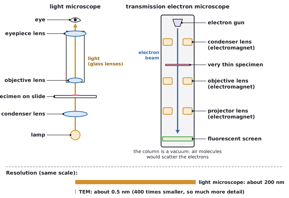 (opens full size in a new tab)
Written and checked against the Edexcel International A Level Biology (YBI11) specification · Updated October 2026