
Contraction and the sliding filament theorySpec 7.11
In short
In the sliding filament theory, an action potential releases calcium ions, which bind troponin and move tropomyosin off the binding sites on actin. Myosin heads attach, pull the actin filaments past the myosin using energy from ATP broken down by ATPase, and the sarcomere shortens as the filaments slide.
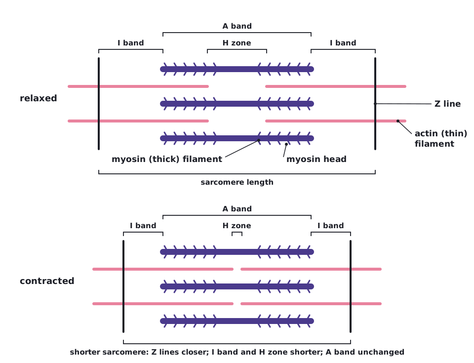
Muscle contracts when the thin actin filaments slide between the thick myosin filaments, pulling the Z lines closer together. The filaments themselves do not shorten.
Roles of the proteins and ions
- Actin: thin filament with binding sites for myosin heads.
- Tropomyosin: fibrous protein that lies along actin and covers the myosin-binding sites when the muscle is relaxed.
- Troponin: a protein attached to tropomyosin that binds Ca²⁺.
- Myosin: thick filament with globular heads that bind actin and act as an ATPase enzyme.
- Ca²⁺: released from the sarcoplasmic reticulum; triggers the change that exposes the binding sites.
- ATP: provides energy for the myosin head to move, and allows it to detach.
- An action potential arrives at the neuromuscular junction and spreads along the sarcolemma and down the T-tubules.
- This causes the sarcoplasmic reticulum to release Ca²⁺ into the sarcoplasm.
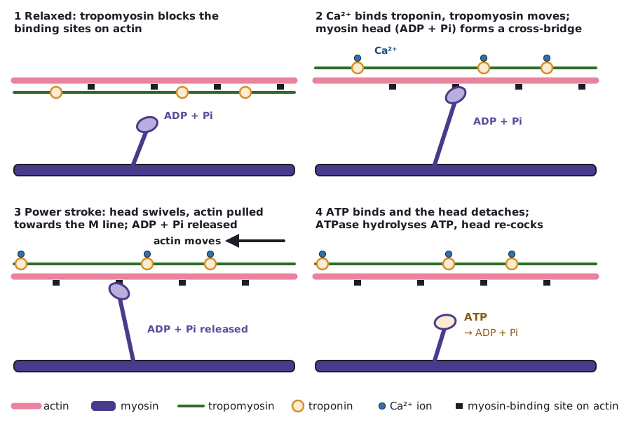
- Ca²⁺ binds to troponin, changing its shape. This pulls tropomyosin away from the myosin-binding sites on actin.
- A myosin head (with ADP and Pi attached) binds to actin, forming an actin–myosin cross-bridge.
- The head changes shape, tilting and pulling the actin filament towards the centre of the sarcomere. ADP and Pi are released. This is the power stroke.
- A new ATP binds to the myosin head, which detaches from the actin.
- Myosin ATPase hydrolyses ATP to ADP and Pi, releasing energy that returns the head to its original position, ready to bind again further along the actin.
- Steps 4 to 7 repeat while Ca²⁺ and ATP are available, and the filaments slide past each other.
When stimulation stops, Ca²⁺ is actively pumped back into the sarcoplasmic reticulum using ATP. Troponin returns to its original shape, tropomyosin covers the binding sites again, and the muscle relaxes.
| Part | Change |
|---|---|
| Sarcomere (Z line to Z line) | Gets shorter |
| I band | Gets shorter |
| H zone | Gets shorter (or disappears) |
| A band | Stays the same length |
ATP is needed to detach the myosin head from actin, as well as to move it. Without ATP the heads stay attached, as in rigor mortis.
 (opens full size in a new tab)
 (opens full size in a new tab)
Quick check
What is the difference between a tendon and a ligament?
Show answer
A tendon joins muscle to bone; a ligament joins bone to bone.
What is an antagonistic muscle pair?
Show answer
Two muscles that have opposite effects at a joint: when one contracts the other relaxes.
Which part of a sarcomere stays the same length when a muscle contracts?
Show answer
The A band.
What does calcium bind to in muscle contraction?
Show answer
Troponin.
Why do fast twitch fibres tire quickly?
Show answer
They use anaerobic respiration, so lactate builds up.
Written and checked against the Edexcel International A Level Biology (YBI11) specification · Updated October 2026