
Prokaryote and eukaryote cell structureSpec A2.2.5, A2.2.6
In short
Prokaryote cells have a cell wall, plasma membrane, cytoplasm, naked DNA in a loop and 70S ribosomes, but no nucleus; Gram-positive eubacteria such as Bacillus and Staphylococcus are the type studied. Eukaryote cells have a compartmentalised cytoplasm with 80S ribosomes, a nucleus with a double membrane and pores, membrane-bound organelles and a cytoskeleton.
Prokaryote cell structure
Prokaryotes have no nucleus. The structure you need is that of Gram-positive eubacteria such as Bacillus and Staphylococcus.
| Component | Description and function |
|---|---|
| Cell wall | A thick layer of peptidoglycan outside the plasma membrane, with no outer membrane beyond it; protects the cell and stops it bursting when water enters by osmosis |
| Plasma membrane | Phospholipid bilayer with proteins; controls the entry and exit of substances |
| Cytoplasm | Mainly water; the site of metabolic reactions; not divided into compartments |
| Naked DNA in a loop | A single circular chromosome, not associated with histones, in a region called the nucleoid |
| 70S ribosomes | Smaller ribosomes, free in the cytoplasm, that carry out protein synthesis |
Prokaryote cell structure varies. Many prokaryotes also have plasmids, flagella, pili or a capsule, but the details of these variations are not required.
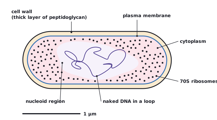 (opens full size in a new tab)
Eukaryote cell structure
- A plasma membrane enclosing a compartmentalised cytoplasm with 80S ribosomes.
- A nucleus with chromosomes made of DNA bound to histones, contained in a double membrane with pores (the nuclear envelope).
- Membrane-bound cytoplasmic organelles: mitochondria, rough and smooth endoplasmic reticulum, the Golgi apparatus and a variety of vesicles or vacuoles, including lysosomes.
- A cytoskeleton of microtubules and microfilaments, which supports the cell, moves organelles and allows some cells to move.
Compartments let enzymes and substrates be concentrated where they are needed, keep potentially harmful enzymes (such as those in lysosomes) away from the rest of the cell, and allow different conditions, such as pH, in different organelles.
| Feature | Prokaryote | Eukaryote |
|---|---|---|
| Nucleus | Absent; DNA in a nucleoid region | Present, with a double membrane and pores |
| DNA | Naked and circular | Linear chromosomes bound to histones |
| Ribosomes | 70S | 80S in the cytoplasm |
| Membrane-bound organelles | Absent | Present: mitochondria, ER, Golgi apparatus, lysosomes |
| Cytoplasm | Not compartmentalised | Compartmentalised |
| Typical size | About 0.5–5 µm | About 10–100 µm |
Do not write that prokaryotes have 'no DNA' or 'no chromosome'. They have DNA, as a single naked loop; what they lack is a nucleus.
Written and checked against the IB Biology SL specification · Updated October 2026