
Micrographs, drawing and annotationSpec A2.2.10, A2.2.11
In short
In light and electron micrographs, cells are identified as prokaryote, plant or animal from features such as the nucleus, cell wall, vacuole and chloroplasts. In electron micrographs you should recognise structures including the mitochondrion, Golgi apparatus and rough and smooth endoplasmic reticulum. Drawings use clear pencil lines and ruled labels, and annotations state each structure's function.
Identifying the cell type
| Cell type | Look for |
|---|---|
| Prokaryote | Small (about 0.5–5 µm); nucleoid region with no nuclear membrane; cell wall; no membrane-bound organelles |
| Plant | Cell wall; large sap vacuole; chloroplasts in green tissue; often a regular, angular shape |
| Animal | No cell wall; no large vacuole; may have microvilli; often an irregular shape |
Structures in electron micrographs
| Structure | Appearance | Function (for annotations) |
|---|---|---|
| Nucleoid region | Paler area of fine fibres in a prokaryote, with no membrane around it | Contains the loop of DNA |
| Prokaryotic cell wall | Dense layer just outside the plasma membrane | Protects the cell and prevents bursting |
| Nucleus | Large and round; double membrane with pores; dark patches of chromatin | Contains the chromosomes; controls the cell through gene expression |
| Chromosomes | Dark, condensed bodies, visible in dividing cells | Carry the genes; condensed so they can be separated in division |
| Mitochondrion | Oval; double membrane, with the inner membrane folded into cristae | Site of aerobic respiration, producing ATP |
| Chloroplast | Double membrane; stacks of thylakoids (grana); may contain starch grains | Site of photosynthesis |
| Sap vacuole | Large, pale area bounded by a single membrane | Stores cell sap; maintains turgor |
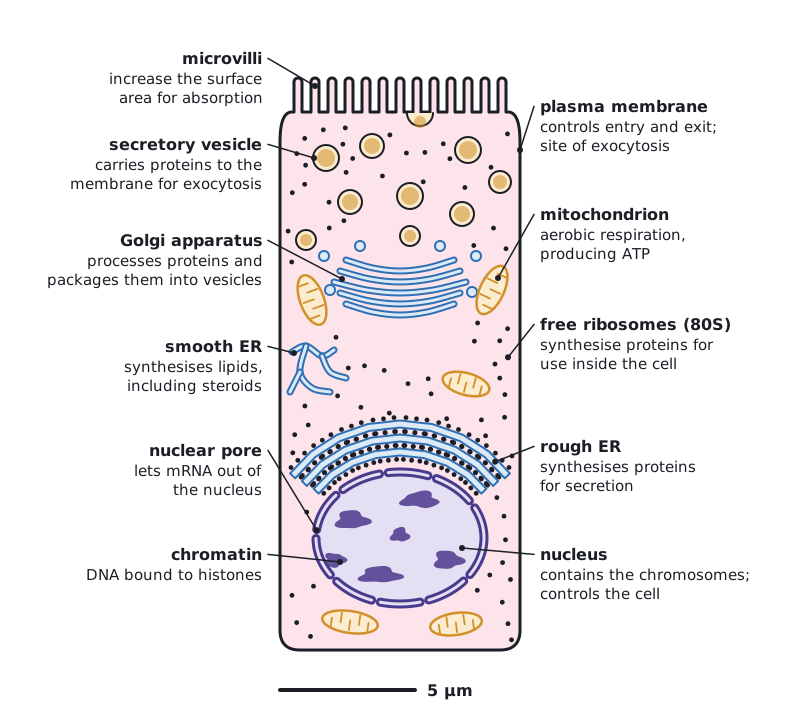
| Golgi apparatus | Stack of curved, flattened membrane sacs with vesicles at the edges | Processes proteins and packages them into vesicles |
| Rough endoplasmic reticulum | Flattened membrane sacs studded with ribosomes | Synthesises proteins for secretion or for membranes |
| Smooth endoplasmic reticulum | Branching tubular membranes without ribosomes | Synthesises lipids, including steroids |
| Ribosomes | Tiny dark granules, free in the cytoplasm or on rough ER | Protein synthesis |
| Cell wall (plant) | Thick layer outside the plasma membrane | Supports the cell and resists turgor pressure |
| Plasma membrane | Thin dark line at the edge of the cytoplasm | Controls the entry and exit of substances |
| Secretory vesicles | Small membrane-bound spheres near the Golgi apparatus or plasma membrane | Carry proteins to the plasma membrane for secretion by exocytosis |
| Microvilli | Finger-like projections of the plasma membrane | Increase the surface area for absorption |
Drawing and annotating from an electron micrograph
- Use a sharp pencil and clear, continuous lines, with no shading or colouring.
- Draw only what is visible, with structures in the correct proportions and positions.
- Label with straight, ruled lines that do not cross and that end exactly on the structure.
- Annotate each labelled structure with its function; this is required, not optional.
- Add a title and a scale bar or magnification.
An annotation is more than a label. 'Mitochondrion: site of aerobic respiration, producing ATP' is an annotation; 'mitochondrion' alone is only a label.
 (opens full size in a new tab)
Quick check
State the three parts of cell theory.
Show answer
All organisms consist of one or more cells; cells are the basic structural unit of life; cells only arise from pre-existing cells.
Why must an eyepiece graticule be calibrated for each objective lens?
Show answer
The length of specimen covered by one eyepiece unit changes with magnification.
What size of ribosome do prokaryotes have?
Show answer
70S.
Which substance makes up fungal cell walls?
Show answer
Chitin.
Written and checked against the IB Biology SL specification · Updated October 2026