
Staining specimensSpec 3.7
In short
Most cell structures are transparent or have little contrast, so specimens are stained. In light microscopy, coloured dyes bind to particular structures and make them visible. In electron microscopy, salts of heavy metals scatter electrons strongly, giving contrast between structures. Different stains can show up different components, so they help to identify structures.
Cells are mostly water and are almost transparent, so structures inside them hardly absorb light and are difficult to see. Staining increases the contrast between structures, and different stains bind to different structures so they can be told apart.
- Light microscopy: coloured dyes absorb light and make structures visible. Examples are methylene blue (stains nuclei and cytoplasm blue), eosin (stains cytoplasm pink), iodine solution (stains starch blue-black) and acetic orcein or toluidine blue (stain DNA/chromosomes dark).
- Differential staining uses stains that bind to different structures, so that two tissues or two parts of a cell are different colours.
- Electron microscopy: the specimen is treated with salts of heavy metals, such as lead or uranium. These scatter electrons, so the stained parts look dark and the rest looks lighter. Different structures take up different amounts of metal, which gives the contrast that allows organelles to be identified.
The reason for staining is always contrast: structures are made visible and distinguishable. Say that, rather than 'to make it clearer'.
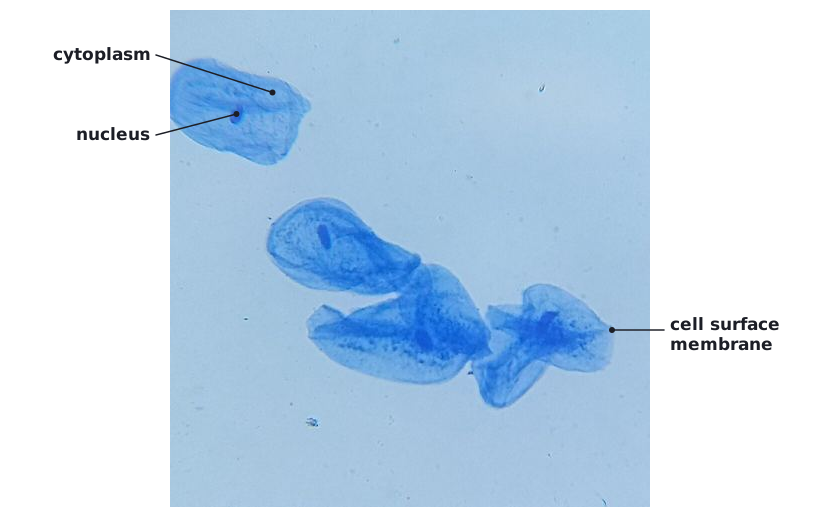 (opens full size in a new tab)
Written and checked against the Edexcel International AS Level Biology (XBI11) specification · Updated October 2026