
The mammalian lungSpec 2.1
In short
Mammalian lungs are adapted for rapid gaseous exchange by millions of alveoli that give a huge surface area, walls only one cell thick, and a dense capillary network. Ventilation and blood flow keep oxygen and carbon dioxide concentration gradients steep, so diffusion is fast.
Air enters the lungs by the trachea, which divides into two bronchi, one to each lung. Each bronchus branches repeatedly into smaller bronchioles, which end in clusters of tiny air sacs called alveoli. Gas exchange happens across the walls of the alveoli.
- Air passes down the trachea, held open by rings of cartilage.
- It moves into a bronchus, then through the many branching bronchioles.
- It reaches the alveoli, where oxygen diffuses into the blood and carbon dioxide diffuses out.
The alveolus and its blood supply
The wall of an alveolus is a single layer of flattened (squamous) epithelial cells. Next to it is the wall of a capillary, which is a single layer of flat endothelial cells. Oxygen therefore diffuses across only two very thin cells (a distance of well under 1 µm in total), from the air in the alveolus into the red blood cells in the capillary.
| Feature | How it helps |
|---|---|
| About 300 to 500 million alveoli in the two human lungs together (a very large number of small sacs) | Gives a very large surface area to volume ratio for gas exchange. |
| Alveolar wall and capillary wall each only one cell thick | Short diffusion distance, so diffusion is rapid (Fick's law: thickness is in the denominator). |
| Dense network of capillaries around each alveolus | Brings a large volume of blood close to the air, and carries oxygen away quickly, which keeps the oxygen concentration gradient steep. |
| Ventilation (breathing in and out) | Brings in fresh air with a high oxygen concentration and removes air with a high carbon dioxide concentration, maintaining both concentration gradients. |
| Blood flow through the capillaries | Continually removes oxygenated blood and delivers blood low in oxygen and high in carbon dioxide, maintaining the gradients. |
| Elastic fibres in the alveolar walls | Stretch as the alveoli fill and recoil when air is breathed out, helping to push air out. |
| Red blood cells squeezed through narrow capillaries | Slows their flow and brings them close to the capillary wall, giving more time for oxygen to be taken up. |
In an 'explain how the lung is adapted' question, link every feature to a consequence for the rate of diffusion. A feature named without its effect ('so diffusion is faster because …') may not earn the mark.
Ventilation does not make gas diffuse faster by itself. It maintains the concentration gradient by replacing the air in the alveoli.
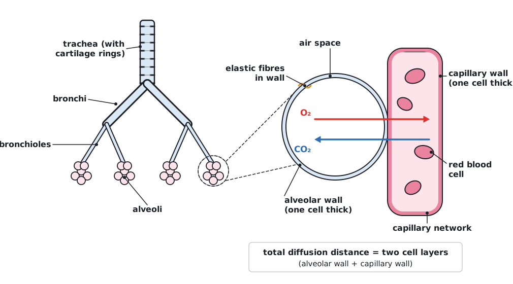 (opens full size in a new tab)
Written and checked against the Edexcel International AS Level Biology (XBI11) specification · Updated October 2026