
Amino acids, peptide bonds and polypeptidesSpec 2.6
In short
An amino acid has a central carbon bonded to an amino group, a carboxyl group, a hydrogen atom and a variable R group. Amino acid monomers are joined by condensation reactions, which release water and form peptide bonds, to make polypeptides. A protein is one or more polypeptides folded into a specific shape.
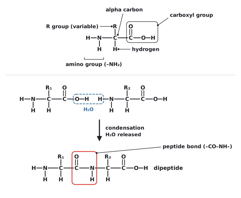
Proteins are polymers made from amino acid monomers. All amino acids share the same basic structure around a central carbon atom: an amino group (–NH₂), a carboxyl group (–COOH), a hydrogen atom, and a variable R group. About 20 different amino acids are used to make proteins, and they differ only in their R group. (You do not need to learn the structures of individual amino acids.)
 (opens full size in a new tab)
Condensation and hydrolysis
Two amino acids join in a condensation reaction. The carboxyl group of one amino acid reacts with the amino group of the next. A molecule of water is released, and a covalent peptide bond (C–N) forms between them, to give a dipeptide. Repeating this builds a chain of many amino acids, called a polypeptide. The reverse reaction, hydrolysis, uses water to break peptide bonds.
A protein is made of one or more polypeptide chains, folded into a precise three-dimensional shape. The sequence of amino acids in each chain is determined by the sequence of bases in a gene.
Recommended additional practical: use a semi-quantitative method to estimate protein concentration with biuret reagent and colour standards. Add sodium hydroxide, then copper(II) sulfate. A purple (lilac) colour shows peptide bonds; the more protein, the deeper the colour. Compare with standards of known concentration or use a colorimeter. Take care: sodium hydroxide is corrosive, so wear eye protection.
The peptide bond is between the carbon of one amino acid's carboxyl group and the nitrogen of the next amino group. A condensation reaction releases water, and does not use it up.
Written and checked against the Edexcel International AS Level Biology (XBI11) specification · Updated October 2026