
Recognising organelles in electron micrographsSpec 3.6
In short
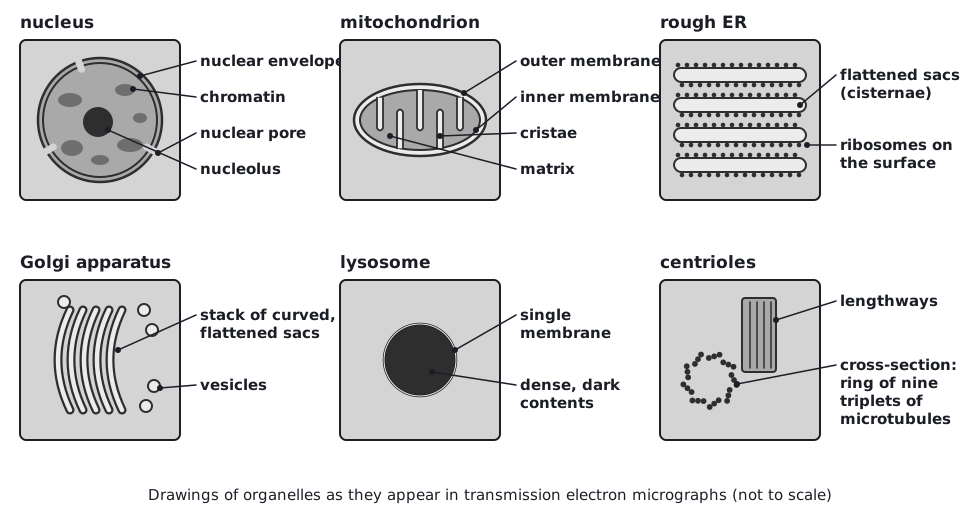
In electron micrographs each organelle has a recognisable appearance. Mitochondria are oval with a folded inner membrane, rough ER is a stack of flattened sacs studded with dots, the Golgi apparatus is a stack of curved sacs with vesicles, and the nucleus has a double envelope with pores and a dark nucleolus.
Electron micrographs are black and white (any colour is added afterwards), and organelles are cut in section, so the shapes you see depend on how they were sliced. Look for these features.
| Organelle | What you see |
|---|---|
| Nucleus | Large, round or oval, with a double membrane broken by pores; dark patches of chromatin; a darker, denser nucleolus |
| Mitochondrion | Small oval or rod shape with a double membrane; the inner membrane folded into cristae, showing as lines or fingers inside |
| Rough ER | Parallel flattened sacs, often continuous with the nuclear envelope, with many tiny black dots (ribosomes) on the outer surface |
| Smooth ER | Branching tubules or sacs with no dots on the surface |
| Golgi apparatus | A stack of curved, flattened sacs, often with small vesicles near the ends |
| Lysosome | Small, round, single membrane, with dense, evenly dark contents |
| Ribosomes | Very small black dots, free or on rER |
| Centrioles | A pair of short cylinders at right angles: one is seen as a ring of microtubules in cross-section and the other lengthways |
Describe what you see rather than guessing: 'double membrane with folded inner membrane' earns the mark for mitochondrion, and you can use it to identify the organelle.
 (opens full size in a new tab)
Quick check
Name three features found in all living cells.
Show answer
Any three of: cell surface membrane, cytoplasm, DNA (genetic material), ribosomes, enzyme-controlled metabolic reactions.
What is the function of the nucleolus?
Show answer
It makes ribosomal RNA and assembles ribosome subunits.
Name the organelle that makes lysosomes.
Show answer
The Golgi apparatus.
How does prokaryotic DNA differ from eukaryotic DNA?
Show answer
It is circular, not associated with histones and not enclosed in a nucleus.
What are the features of a mitochondrion in an electron micrograph?
Show answer
Oval shape, a double membrane and an inner membrane folded into cristae.
Written and checked against the Edexcel International AS Level Biology (XBI11) specification · Updated October 2026