
Structure of the mammalian heartSpec 1.8
In short
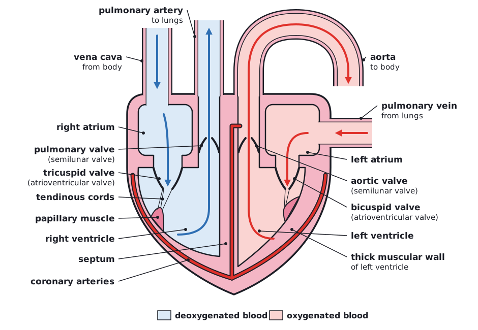
The mammalian heart has four chambers: two thin-walled atria that receive blood and two thicker-walled ventricles that pump it out. The septum separates oxygenated from deoxygenated blood. Atrioventricular and semilunar valves prevent backflow, and the left ventricle has the thickest wall because it pumps blood at high pressure around the whole body.
 (opens full size in a new tab)
| Vessel | Carries blood | From | To |
|---|---|---|---|
| Vena cava | Deoxygenated | Body | Right atrium |
| Pulmonary artery | Deoxygenated | Right ventricle | Lungs |
| Pulmonary vein | Oxygenated | Lungs | Left atrium |
| Aorta | Oxygenated | Left ventricle | Body |
| Coronary arteries | Oxygenated | Base of the aorta | Heart muscle (cardiac muscle) |
- The atria have thin walls because they only pump blood a short distance into the ventricles.
- The ventricles have thicker, muscular walls to pump blood out of the heart. The left ventricle has the thickest wall because it pumps blood to the whole body at high pressure. The right ventricle pumps blood only to the nearby lungs at lower pressure, which protects the lung capillaries.
- The septum separates the left and right sides, so oxygenated and deoxygenated blood do not mix.
- Atrioventricular valves (tricuspid on the right, bicuspid on the left) stop blood flowing back from the ventricles into the atria. They are held in position by tendinous cords attached to papillary muscles, so that they are not pushed inside out when the ventricles contract.
- Semilunar valves at the start of the aorta and pulmonary artery stop blood flowing back from the arteries into the ventricles.
- The coronary arteries supply the cardiac muscle with oxygen and glucose. Blockage of a coronary artery causes a heart attack.
Recommended additional practical (not a core practical): investigate the structure of a mammalian heart by dissection. Cut across the ventricles to compare wall thickness, find the valves and tendinous cords, and identify the vessels. Wear gloves and eye protection, use a sharp scalpel on a dissecting board, cut away from your hand and disinfect surfaces afterwards.
Written and checked against the Edexcel International AS Level Biology (XBI11) specification · Updated October 2026