

The structure of the thoraxSpec 2.46
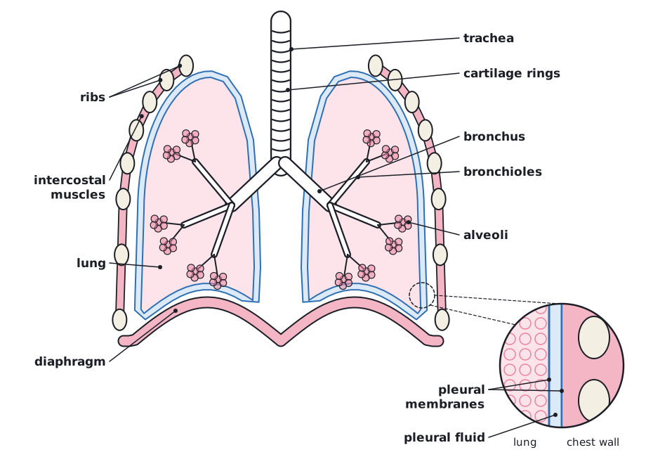
The thorax is the chest. It contains the lungs and the heart, and it is protected by the ribs. Air enters through the nose or mouth and passes along the airways to the alveoli.
| Structure | Description and function |
|---|---|
| Ribs | Bones that form a cage and protect the lungs and heart. They can move up and out, and down and in. |
| Intercostal muscles | Muscles between the ribs. They move the rib cage. |
| Diaphragm | A sheet of muscle under the lungs that separates the thorax from the abdomen. |
| Trachea | The windpipe. It carries air to the lungs. Rings of cartilage keep it open. |
| Bronchi (one is a bronchus) | Two tubes that branch from the trachea, one to each lung. |
| Bronchioles | Small tubes that branch from the bronchi and carry air to the alveoli. |
| Alveoli | Tiny air sacs at the ends of the bronchioles, where gas exchange happens. |
| Pleural membranes | Two thin membranes around the lungs and lining the chest wall, with a thin layer of fluid between them. The fluid reduces friction when the lungs move. |
The path of air when you breathe in is: nose or mouth, trachea, bronchi, bronchioles, alveoli.
Exam tip:
Learn the order of the airways. Marks are often lost by writing bronchi and bronchioles the wrong way round: the bronchi are the larger tubes.
 (opens full size in a new tab)