

Structure and function of the nervous systemSpec 4.5.2.1
The nervous system enables humans to react to their surroundings and to coordinate their behaviour.
Information from receptors passes along cells called neurones as electrical impulses to the central nervous system (CNS). The CNS is the brain and spinal cord. The CNS coordinates the response of effectors, which may be muscles contracting or glands secreting hormones.
- Stimulus (a change in the environment)
- Receptor detects the stimulus
- Coordinator (the CNS) processes the information
- Effector (a muscle or a gland)
- Response
How the structure suits the function
| Part | Adaptation | Why it helps |
|---|---|---|
| Receptors | Each type is sensitive to one kind of stimulus | The body only picks up the changes it needs to respond to |
| Neurones | Long cells that carry electrical impulses | Impulses travel quickly over long distances between receptors, the CNS and effectors |
| CNS | The brain and spinal cord receive information and send out a coordinated response | The right effectors respond together |
| Effectors | Muscles contract; glands secrete hormones | They carry out the response |
Reflex actions and the reflex arcSpec 4.5.2.1
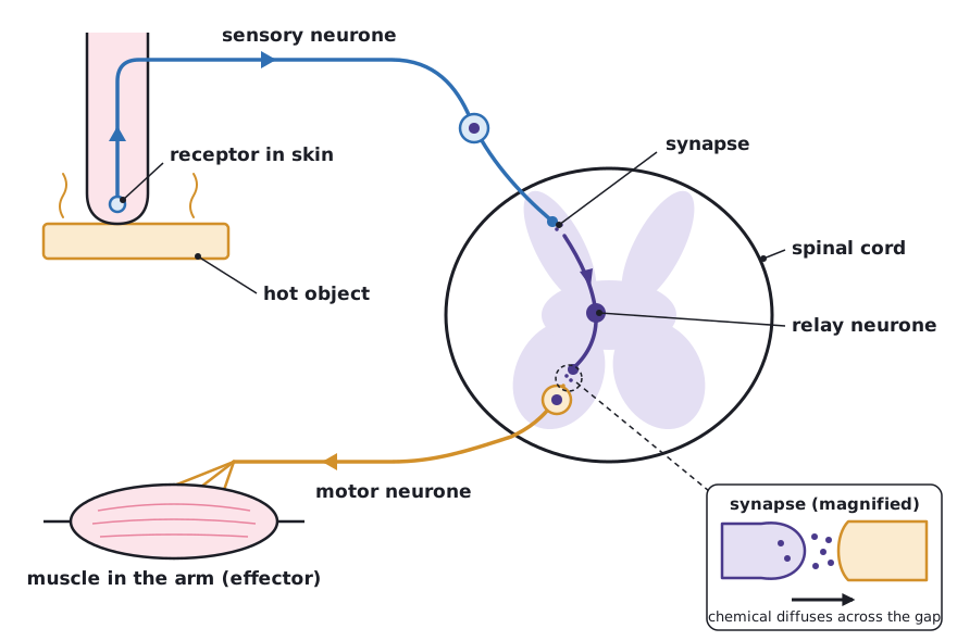
Reflex actions are automatic and rapid. They do not involve the conscious part of the brain. Reflexes are important because they protect the body from harm, for example pulling your hand away from a hot object before you have time to think.
- A receptor detects the stimulus and an impulse starts in the sensory neurone.
- The sensory neurone carries the impulse to the CNS.
- At a synapse, chemicals diffuse across the gap and start an impulse in the relay neurone.
- The relay neurone passes the impulse on, through another synapse, to the motor neurone.
- The motor neurone carries the impulse to the effector.
- The effector (a muscle or gland) produces the response.
| Structure | Function |
|---|---|
| Receptor | Detects the stimulus |
| Sensory neurone | Carries impulses from the receptor to the CNS |
| Relay neurone | Carries impulses within the CNS, from the sensory neurone to the motor neurone |
| Synapse | A gap between two neurones. A chemical diffuses across the gap and starts an impulse in the next neurone |
| Motor neurone | Carries impulses from the CNS to the effector |
| Effector | A muscle (contracts) or a gland (secretes) that produces the response |
 Tap to enlarge
Required practical: reaction timeSpec 4.5.2.1
Reaction time is the time taken to respond to a stimulus. A common method uses a ruler.
- The volunteer sits with their forearm resting on the edge of a table and their hand over the edge.
- A partner holds a ruler vertically so that the zero mark is level with the volunteer’s thumb and finger, which are about to grip it.
- Without warning, the partner drops the ruler.
- The volunteer catches it as fast as possible. Read the distance on the ruler at the top of their thumb.
- Repeat several times and calculate a mean for each condition.
- Independent variable: the factor you change, for example caffeine, a distraction, practice or which hand is used.
- Dependent variable: reaction time, shown by the distance the ruler falls (a shorter distance means a faster reaction), or by a time from a computer test.
- Control variables: the same volunteer, the same hand, the same ruler, the same drop height, and the same method of reading the result.
- Safety: the risk is low. Make sure nobody is pressured into taking a drink or doing anything they are not comfortable with.
Repeating the test and taking a mean makes the results more reliable. Ignore any anomalous result when you calculate the mean.
From numbers to a graph
A student measured reaction time (in s) five times: 0.31, 0.29, 0.33, 0.30, 0.32. Calculate the mean, then say how the means for two conditions would be shown on a bar chart.
- Add the five results: 0.31 + 0.29 + 0.33 + 0.30 + 0.32 = 1.55
- Divide by the number of results: 1.55 ÷ 5 = 0.31
- Draw one bar per condition, labelled on the x-axis, with bar height equal to the mean reaction time on the y-axis (in s).
Answer: Mean reaction time = 0.31 s.
The brainSpec 4.5.2.2Triple only
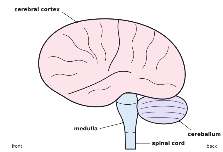
The brain controls complex behaviour. It is made of billions of interconnected neurones and has different regions that carry out different functions.
| Region | Function |
|---|---|
| Cerebral cortex | Consciousness, intelligence, memory and language |
| Cerebellum | Coordinates muscular activity and balance |
| Medulla | Controls unconscious activities such as heart rate and breathing |
 Tap to enlarge
Investigating the brain and treating brain disorders
Neuroscientists have mapped regions of the brain to particular functions in three main ways.
- Studying patients with brain damage and seeing which abilities are lost.
- Electrically stimulating different parts of the brain and seeing what happens.
- Using MRI scanning techniques.
The complexity and delicacy of the brain makes investigating and treating brain disorders very difficult. It is made of billions of interconnected neurones, and its tissue is easily damaged during investigation or treatment. Any procedure on the brain or nervous system has benefits and risks that must be weighed up.
The eye: structures and their functionsSpec 4.5.2.3Triple only
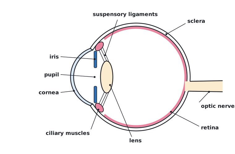
The eye is a sense organ containing receptors sensitive to light intensity and colour.
| Structure | How its structure relates to its function |
|---|---|
| Retina | Contains the light receptors. It is where the image is focused and it detects light intensity and colour |
| Optic nerve | Carries impulses from the receptors in the retina to the brain |
| Sclera | The tough white outer layer. It protects the eye and keeps its shape |
| Cornea | Transparent front surface. It lets light in and refracts (bends) the light rays |
| Iris | Muscles that control the size of the pupil and so how much light enters the eye |
| Ciliary muscles | A ring of muscle around the lens that contracts or relaxes to change the lens shape |
| Suspensory ligaments | Hold the lens in place and connect it to the ciliary muscles. Their tension changes the lens shape |
 Tap to enlarge
Adaptation to dim light
The iris controls how much light reaches the retina. In dim light the pupil gets wider so more light enters. In bright light the pupil gets narrower so less light enters, which protects the retina.
AccommodationSpec 4.5.2.3Triple only
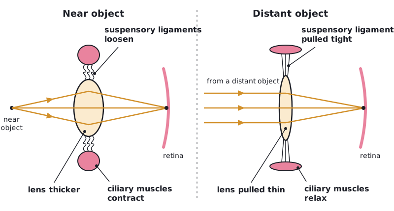
- Accommodation
- The process of changing the shape of the lens to focus on near or distant objects.
| Near object | Distant object | |
|---|---|---|
| Ciliary muscles | Contract | Relax |
| Suspensory ligaments | Loosen | Pulled tight |
| Lens shape | Thicker | Pulled thin |
| Refraction of light rays | Strongly | Only slightly |
 Tap to enlarge
Defects of the eyeSpec 4.5.2.3Triple only
Two common defects of the eye are myopia (short sightedness) and hyperopia (long sightedness). In both, rays of light do not focus on the retina.
| Myopia (short sightedness) | Hyperopia (long sightedness) | |
|---|---|---|
| Effect | Distant objects look blurred | Near objects look blurred |
| Where light rays focus | In front of the retina | Behind the retina |
| Correction with spectacle lenses | Concave lens, which spreads the light rays slightly before they enter the eye | Convex lens, which bends the light rays inwards before they enter the eye |
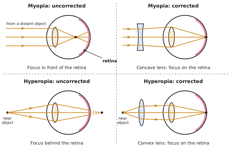 Tap to enlarge
Generally these defects are treated with spectacle lenses which refract the light rays so that they do focus on the retina. New technologies now include:
- hard and soft contact lenses
- laser surgery to change the shape of the cornea
- a replacement lens in the eye.
Control of body temperatureSpec 4.5.2.4Triple only
Body temperature is monitored and controlled by the thermoregulatory centre in the brain. It contains receptors sensitive to the temperature of the blood. The skin also contains temperature receptors and sends nervous impulses to the thermoregulatory centre.
| If body temperature is… | Responses |
|---|---|
| Too high | Blood vessels dilate (vasodilation) and sweat is produced from the sweat glands. Both cause a transfer of energy from the skin to the environment |
| Too low | Blood vessels constrict (vasoconstriction), sweating stops and skeletal muscles contract (shiver) |
Explaining how the mechanisms work
- Vasodilation: more blood flows through the vessels near the skin surface, so more energy is transferred from the blood to the surroundings.
- Sweating: the sweat evaporates from the skin surface, which transfers energy from the skin to the environment and cools the body.
- Vasoconstriction: less blood flows near the skin surface, so less energy is transferred to the surroundings.
- Shivering: skeletal muscles contract rapidly, and the respiration in the muscle cells releases energy that warms the body.
- Sweating stops, so less energy is lost by evaporation.
Quick check
What is the CNS?
Show answer
The central nervous system: the brain and spinal cord.
Put these in order: motor neurone, sensory neurone, relay neurone.
Show answer
Sensory neurone, relay neurone, motor neurone.
Why are reflex actions important?
Show answer
They are automatic and rapid, protecting the body from harm without involving the conscious part of the brain.
Triple only What happens to the ciliary muscles and lens when focusing on a near object?
Show answer
The ciliary muscles contract, the suspensory ligaments loosen and the lens becomes thicker.
Triple only Name the type of lens that corrects myopia.
Show answer
A concave lens.