

AntibioticsSpec 5.16
Antibiotics are medicines that treat bacterial infections. They work by inhibiting cell processes in the bacterium. They do not harm the host organism's own cells, because the host's cells work differently.
Antibiotics can only be used to treat bacterial infections. Viruses are not cells. They live and reproduce inside host cells, so the cell processes that antibiotics inhibit are not present in a virus. Antibiotics therefore do not work on viral diseases such as HIV or Ebola.
Aseptic techniquesSpec 5.17BTriple only
To grow (culture) microorganisms safely and get reliable results, you must stop unwanted microorganisms getting in and stop your culture escaping. This is called aseptic technique.
- Use an autoclave to prepare the sterile growth medium (agar jelly) and the petri dishes. An autoclave sterilises equipment using high-pressure steam, which kills microorganisms already present.
- Use a sterile inoculating loop to transfer microorganisms. The loop is sterilised by heating in a flame until red hot, then cooled before use.
- Keep petri dishes and culture vials covered as much as possible. Only lift the lid briefly when transferring microorganisms.
- After inoculating, fix the lid on with tape and incubate the dish upside down.
- Wash your hands and wipe the bench with disinfectant before and after.
Core practical: antiseptics, antibiotics and plant extractsSpec 5.18BTriple only
The idea is to find out which substance is best at stopping bacteria growing. Bacteria are grown on agar jelly in a petri dish, and small paper discs soaked in different substances are placed on top.
- Using aseptic technique, spread a sample of bacteria evenly over the surface of a sterile agar plate to make a bacterial lawn.
- Using sterile forceps, place paper discs soaked in the different antiseptics, antibiotics or plant extracts on the agar, spaced apart.
- Place a control disc soaked in sterile water on the agar.
- Put the lid on and tape it in place. Label the base and incubate the dish upside down at 25 °C for about 48 hours.
- Measure the diameter of the clear zone (inhibition zone) around each disc and calculate its area.
| Type of variable | In this investigation |
|---|---|
| Independent | The type of antiseptic, antibiotic or plant extract (or its concentration) |
| Dependent | The size of the clear zone |
| Control | Type and volume of bacteria; type and volume of agar; size of disc and volume of liquid on it; temperature and time of incubation |
- Results: where the substance stops bacteria growing, a clear zone appears around the disc. The larger the clear zone, the more effective the substance. The water control disc should have no clear zone.
- Why 25 °C: a lower temperature makes it less likely that harmful pathogens that grow best at body temperature will grow.
- Why the dish is taped: to stop microorganisms escaping or entering; do not seal it fully, so that oxygen can enter.
- Safety: wear eye protection; do not open dishes after incubation; wash hands; disinfect the bench; sterilise used dishes in an autoclave; take care with flammable chemicals such as ethanol near a flame.
Calculating cross-sectional areasSpec 5.19BTriple only
Clear zones and bacterial colonies are roughly circular, so their area is calculated using the equation for the area of a circle. A larger area means a bigger effect (for a clear zone) or more growth (for a culture).
The radius, r, is half the diameter. Measure the diameter across the middle of the zone, halve it, then square it and multiply by π.
Area of a clear zone
The clear zone around a disc soaked in antiseptic has a diameter of 14 mm. Calculate the area of the clear zone. Use π = 3.14.
- radius = 14 ÷ 2 = 7 mm
- area = πr² = 3.14 × 7 × 7
- area = 3.14 × 49 = 153.86 mm²
Answer: About 154 mm²
Developing new medicinesSpec 5.20
Developing a new medicine, including a new antibiotic, takes many stages. Each stage checks that the medicine is safe and that it works.
- Discovery: finding a chemical that might treat the disease. These may come from natural sources, such as plants or microorganisms, or be designed in a laboratory.
- Development: the chemical is studied and improved, and a way to make it as a medicine is worked out.
- Preclinical testing: the medicine is tested in the laboratory on cells and tissues, and then on live animals, to find out if it is toxic and whether it works.
- Clinical testing: the medicine is tested on people. It is first given to a small number of healthy volunteers to check it is safe, then to patients to check that it works and to find the right dose.
Only a medicine that passes all the stages can be used to treat patients. The testing protects people from harmful or ineffective medicines.
Producing monoclonal antibodiesSpec 5.21BTriple onlyHigher tier
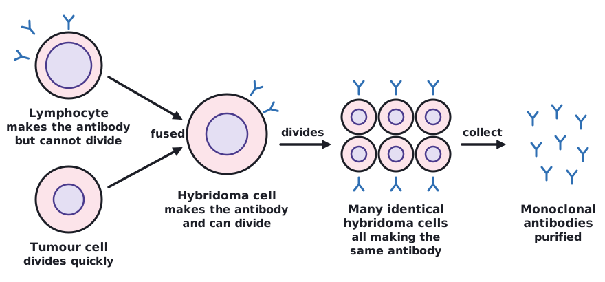
Monoclonal antibodies are identical antibodies, all made from one type of cell, that bind to only one specific antigen.
- An animal is exposed to the antigen, so it makes lymphocytes that produce the desired antibody.
- These lymphocytes are collected. They make the right antibody but they do not divide.
- They are fused with a tumour cell, which divides very quickly. This makes a hybridoma cell.
- The hybridoma cells divide and so make many identical cells. As they divide, the hybridoma cells produce antibodies.
- The antibodies are collected and purified.
A hybridoma cell has the properties of both its parent cells: it makes the desired antibody (from the lymphocyte) and it can divide again and again (from the tumour cell).
 Tap to enlarge
Uses of monoclonal antibodiesSpec 5.22BTriple onlyHigher tier
Monoclonal antibodies bind to one specific antigen only, so they can be used to find or target particular molecules or cells.
- Pregnancy testing: a test stick contains monoclonal antibodies that bind to a hormone found in the urine of pregnant women. When the hormone is present, it binds to the antibodies and a coloured line appears, so the test is positive.
- Diagnosis: monoclonal antibodies carrying a label can be given to a patient. They bind to a specific antigen, so the label shows the position of blood clots or cancer cells.
- Treatment: a drug can be attached to a monoclonal antibody that binds to antigens on cancer cells. The antibody carries the drug directly to the cancer cells.
Advantages over drug and radiotherapy treatments
Drugs and radiotherapy can damage healthy cells as well as cancer cells, which causes side effects. Monoclonal antibodies target specific cells, so the cancer cells are attacked and fewer healthy cells are damaged.
Quick check
Why can antibiotics not be used to treat viral infections?
Show answer
Antibiotics inhibit cell processes in bacteria; viruses are not cells and reproduce inside host cells.
Triple only: why is an inoculating loop heated in a flame before use?
Show answer
To sterilise it by killing any microorganisms on it.
Triple only: in the core practical, what does a larger clear zone show?
Show answer
The substance is more effective at stopping bacteria growing.
Name the four stages in developing a new medicine in order.
Show answer
Discovery, development, preclinical testing, clinical testing.
Triple only, Higher tier: what is a hybridoma cell?
Show answer
A cell made by fusing an antibody-producing lymphocyte with a tumour cell; it divides and produces antibodies.