

The digestive systemSpec 4.2.2.1
The digestive system is an example of an organ system in which several organs work together to digest and absorb food.
Food contains large molecules of carbohydrate, protein and lipid. These are too big to pass into the blood. Digestive enzymes convert food into small soluble molecules that can be absorbed into the bloodstream.
| Organ | Role |
|---|---|
| Salivary glands | Make amylase, which mixes with food in the mouth |
| Stomach | Churns food. Makes protease and hydrochloric acid |
| Liver | Makes bile |
| Gall bladder | Stores bile |
| Pancreas | Makes amylase, protease and lipase, and releases them into the small intestine |
| Small intestine | Makes amylase, protease and lipase. Digestion is finished here and the small soluble molecules are absorbed into the blood |
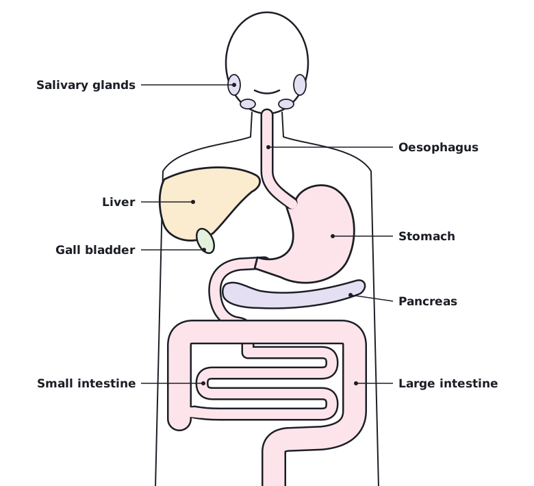 Tap to enlarge
EnzymesSpec 4.2.2.1
Enzymes catalyse specific reactions in living organisms. A catalyst speeds up a reaction and is not used up. Enzymes are large protein molecules, and each one has a special shape.
Each enzyme has an active site. The shape of the active site is what makes the enzyme specific: it only works on one type of molecule, called the substrate.
The lock and key theory
The ‘lock and key theory’ is a simplified model of how enzymes work.
- The substrate collides with the enzyme and fits into the active site, like a key into a lock. The shapes are complementary.
- The enzyme catalyses the reaction while the substrate is in the active site.
- The products leave the active site. The enzyme is unchanged and can be used again.
Scientists also use other models to explain enzyme action. If a question describes a different model, use the information given in the question.
Temperature and pH
As temperature rises, the reaction gets faster because the particles move faster and collide more often. Each enzyme has an optimum temperature where its rate is highest.
If the temperature is too high, or the pH is too acidic or too alkaline, the shape of the active site changes. The substrate no longer fits. The enzyme is denatured, and it cannot be changed back.
| Condition | Effect on rate |
|---|---|
| Temperature below the optimum | Rate increases as temperature rises |
| Optimum temperature | Rate is highest |
| Temperature above the optimum | Rate falls quickly because the enzyme is denatured |
| Optimum pH | Rate is highest. Different enzymes have different optimum pH values |
| pH far from the optimum | Rate falls because the enzyme is denatured |
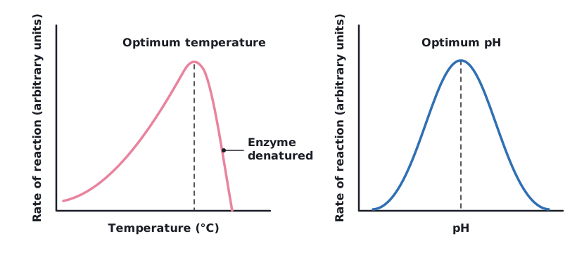 Tap to enlarge
Rate calculations
When the end point is a colour change, such as the loss of starch, you can use rate = 1 ÷ time taken. A shorter time means a faster rate.
Rate of an enzyme-catalysed reaction
An enzyme makes 12 cm³ of product in 3 minutes. Calculate the rate of reaction in cm³ per minute.
- Use rate = amount of product ÷ time.
- Rate = 12 ÷ 3.
Answer: 4 cm³/min
Amylase, proteases and lipasesSpec 4.2.2.1
Three types of digestive enzyme break the three main food molecules into small, soluble molecules. You need to recall where each is made and what it does.
| Enzyme | Breaks down | Into | Made in |
|---|---|---|---|
| Amylase (a carbohydrase) | Starch | Simple sugars | Salivary glands, pancreas, small intestine |
| Protease | Proteins | Amino acids | Stomach, pancreas, small intestine |
| Lipase | Lipids (fats) | Glycerol and fatty acids | Pancreas, small intestine |
Carbohydrases break down carbohydrates to simple sugars. Amylase is a carbohydrase which breaks down starch.
You only need simple word equations, not chemical symbol equations.
The products of digestion are used to build new carbohydrates, lipids and proteins. Some glucose is used in respiration.
BileSpec 4.2.2.1
Bile is made in the liver and stored in the gall bladder. It is released into the small intestine.
Bile does two jobs:
- It is alkaline, so it neutralises hydrochloric acid that comes from the stomach. This gives the enzymes in the small intestine the alkaline conditions they work best in.
- It emulsifies fat, breaking it into small droplets. This increases the surface area of the fat.
The alkaline conditions and the large surface area increase the rate of fat breakdown by lipase.
Required practical: food testsSpec 4.2.2.1
You can use qualitative reagents to test for a range of carbohydrates, lipids and proteins. Qualitative means the test tells you whether a food molecule is present, not how much.
- Prepare a food sample: grind a small amount of food with water and filter it or let it settle. Test the liquid.
- Add the test reagent to a sample of the food solution.
- Look for the colour change and compare with the table below.
| Food molecule | Test | Positive result |
|---|---|---|
| Sugars | Benedict's test: add Benedict's solution (blue) and heat in a hot water bath | Blue changes to green, yellow, orange or brick-red |
| Starch | Iodine test: add iodine solution (orange-brown) | Orange-brown changes to blue-black |
| Protein | Biuret test: add Biuret reagent (blue) | Blue changes to purple (lilac) |
| Lipids | Sudan III test: add a few drops of Sudan III stain to the sample in water and shake | A red-stained oily layer forms on top of the water |
Some schools use the ethanol emulsion test for lipids instead: shake the sample with ethanol, then pour it into water. A cloudy white emulsion shows that lipid is present.
The more the Benedict's colour moves towards brick-red, the more sugar there is. If the colour stays blue, no sugar is present.
Required practical: the effect of pH on amylaseSpec 4.2.2.1
This practical investigates how pH affects the rate of reaction of the enzyme amylase. You use a continuous sampling technique to find the time taken to completely digest a starch solution at a range of pH values.
- Put a drop of iodine solution into each well of a spotting tile.
- Add starch solution and a buffer solution of a known pH to a test tube. Add amylase solution.
- Place the tube in a water bath (or use an electric heater) so the temperature stays constant. Start a stopwatch.
- Every 30 seconds, take a sample from the tube and add it to a fresh drop of iodine.
- Stop when the iodine stays orange-brown. All the starch has been digested. Record the time taken.
- Repeat at a range of pH values, using a different buffer each time.
| Type | Variable |
|---|---|
| Independent | pH of the buffer |
| Dependent | Time taken for the starch to be completely digested (rate = 1 ÷ time) |
| Control | Temperature, volume and concentration of starch and amylase |
The shorter the time, the faster the rate. The rate is highest at the optimum pH. Away from the optimum, amylase is denatured and the starch takes much longer to digest, or is not digested at all.
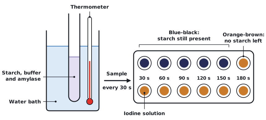 Tap to enlarge
The heart and the double circulatory systemSpec 4.2.2.2
The heart is an organ that pumps blood around the body in a double circulatory system. There are two separate loops, and blood passes through the heart twice on each complete circuit.
- The right ventricle pumps blood to the lungs, where gas exchange takes place.
- The left ventricle pumps blood around the rest of the body.
Blood returns from the body to the right atrium and from the lungs to the left atrium. The ventricles have thick muscular walls. The left ventricle has a thicker wall than the right, because it has to pump blood all the way around the body at a higher pressure. Valves in the heart stop blood flowing backwards. You do not need to know the names of the valves.
| Vessel | Carries blood | Oxygen content |
|---|---|---|
| Vena cava | From the body to the right atrium | Low (deoxygenated) |
| Pulmonary artery | From the right ventricle to the lungs | Low (deoxygenated) |
| Pulmonary vein | From the lungs to the left atrium | High (oxygenated) |
| Aorta | From the left ventricle to the body | High (oxygenated) |
| Coronary arteries | Over the surface of the heart, supplying the heart muscle | High (oxygenated) |
- Deoxygenated blood from the body enters the right atrium through the vena cava.
- It passes into the right ventricle, which pumps it to the lungs through the pulmonary artery.
- In the lungs the blood picks up oxygen and loses carbon dioxide.
- Oxygenated blood returns through the pulmonary vein to the left atrium.
- It passes into the left ventricle, which pumps it around the body through the aorta.
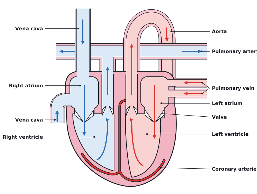 Tap to enlarge
Controlling the heart rateSpec 4.2.2.2
The natural resting heart rate is controlled by a group of cells located in the right atrium that act as a pacemaker.
Sometimes the heart rate is irregular. Artificial pacemakers are electrical devices used to correct irregularities in the heart rate.
The lungs and gas exchangeSpec 4.2.2.2
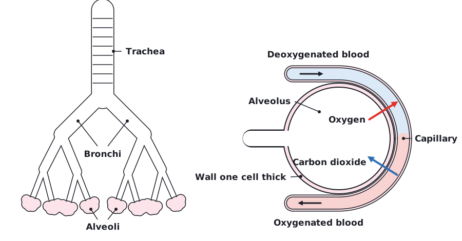
You need to know the structure of the lungs and how they are adapted for gaseous exchange. Knowledge of the lungs is restricted to the trachea, bronchi, alveoli and the capillary network surrounding the alveoli.
- Air passes down the trachea.
- The trachea divides into two bronchi, one to each lung.
- The bronchi branch into smaller tubes that end in alveoli (air sacs).
- At the alveoli, oxygen diffuses into the blood in the surrounding capillaries and carbon dioxide diffuses out of the blood into the air.
Blood arriving at the lungs has a low concentration of oxygen and a high concentration of carbon dioxide. Oxygen therefore diffuses from the air in the alveoli into the blood, and carbon dioxide diffuses the other way.
| Adaptation | Why it helps |
|---|---|
| Millions of alveoli | Gives a very large surface area for diffusion |
| Walls one cell thick | Short diffusion distance, so gases cross quickly |
| Surrounded by a network of capillaries (a good blood supply) | Blood carries oxygen away and brings carbon dioxide, which keeps a steep concentration gradient |
| Constantly ventilated by breathing | Fresh air keeps the oxygen concentration high in the alveoli, which also keeps a steep concentration gradient |
 Tap to enlarge
Blood vesselsSpec 4.2.2.2
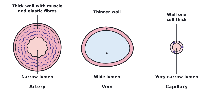
The body contains three different types of blood vessel: arteries, veins and capillaries. Their structures are related to their functions.
| Artery | Vein | Capillary | |
|---|---|---|---|
| Function | Carries blood away from the heart at high pressure | Carries blood back to the heart at low pressure | Exchanges substances with body cells |
| Wall | Thick, with muscle and elastic fibres | Thinner walls | One cell thick, so substances pass through easily |
| Lumen (inside space) | Narrow compared with the thickness of the wall | Wide | Very narrow |
| Valves | No | Yes, to stop backflow | No |
Arteries have thick, strong walls because the blood is at high pressure. The elastic fibres let the wall stretch and spring back. The muscle strengthens the wall.
Veins have a wide lumen and valves. The blood is at low pressure, so the valves stop it flowing backwards.
Capillaries are tiny and have very thin walls. This gives a short diffusion distance, so oxygen and nutrients can pass to the cells and carbon dioxide and waste can pass back into the blood. Capillary walls are permeable.
Blood flow rate
Calculating blood flow
300 cm³ of blood flows through an artery in 4 minutes. Calculate the rate of blood flow in cm³ per minute.
- Rate = volume ÷ time.
- Rate = 300 ÷ 4.
Answer: 75 cm³/min
 Tap to enlarge
BloodSpec 4.2.2.3
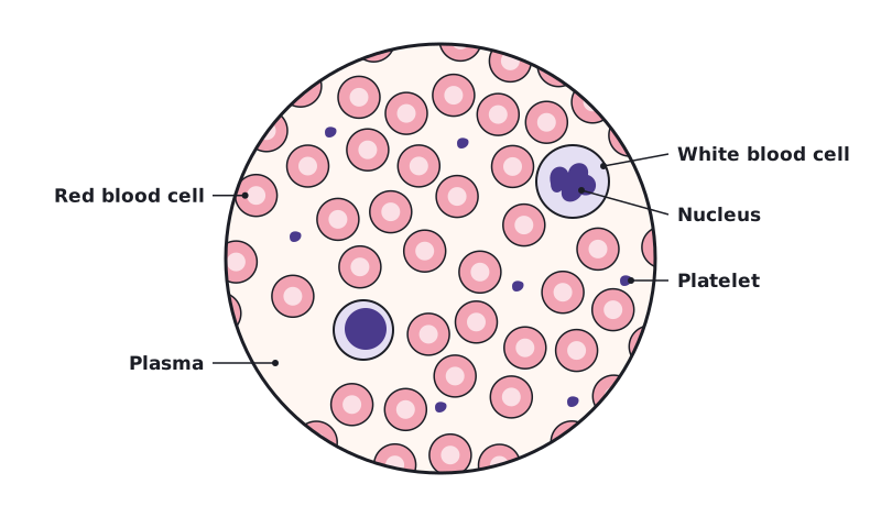
Blood is a tissue consisting of plasma, in which the red blood cells, white blood cells and platelets are suspended.
| Component | Function | Adaptation |
|---|---|---|
| Plasma | Transports carbon dioxide from the organs to the lungs, soluble products of digestion from the small intestine to other organs, and urea from the liver to the kidneys. Also transports hormones | A liquid, so substances dissolve in it |
| Red blood cells | Carry oxygen from the lungs to all the cells of the body | Biconcave disc shape gives a large surface area. No nucleus, so more room for haemoglobin. Contain haemoglobin, which binds to oxygen |
| White blood cells | Defend the body against pathogens. Some engulf pathogens (phagocytosis); others produce antibodies and antitoxins | Have a nucleus. Phagocytes can change shape to engulf pathogens |
| Platelets | Help the blood to clot at a wound | Small fragments of cells with no nucleus |
In the lungs, haemoglobin combines with oxygen to form oxyhaemoglobin. In the body tissues the oxyhaemoglobin releases the oxygen.
Recognising blood cells
You may need to identify cells in a photograph or diagram.
- Red blood cells are the most numerous. They are small, round and have no nucleus, and look paler in the middle.
- White blood cells are larger and have a clear nucleus, often stained dark purple.
- Platelets are tiny fragments, much smaller than the other cells.
Blood products, such as donated blood used in a transfusion, can save lives. There are risks, for example infection can be passed on from the donor, so donated blood has to be checked and matched to the patient.
 Tap to enlarge
Coronary heart diseaseSpec 4.2.2.4
Coronary heart disease (CHD) is a non-communicable disease: it is not caused by a pathogen and cannot be passed from person to person.
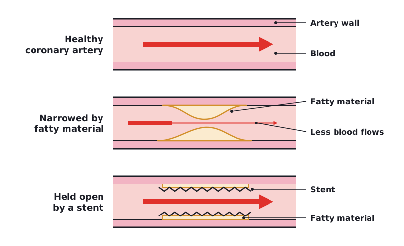
In CHD, layers of fatty material build up inside the coronary arteries, narrowing them. This reduces the flow of blood through the coronary arteries, resulting in a lack of oxygen for the heart muscle.
- Stents are used to keep the coronary arteries open.
- Statins are widely used to reduce blood cholesterol levels, which slows down the rate of fatty material deposit.
- Fatty material builds up inside a coronary artery.
- The artery narrows, so less blood flows through it.
- The heart muscle receives less oxygen.
 Tap to enlarge
Treating cardiovascular diseaseSpec 4.2.2.4
Faulty heart valves
In some people heart valves may become faulty. A valve may be prevented from opening fully, or it might develop a leak.
- If a valve does not open fully, less blood can flow through, so the heart has to work harder and the body may receive less oxygen.
- If a valve leaks, blood flows backwards, so the heart pumps less blood forward with each beat.
Faulty heart valves can be replaced using biological or mechanical valves.
Heart failure
In the case of heart failure, a donor heart, or heart and lungs, can be transplanted. Artificial hearts are occasionally used to keep patients alive whilst waiting for a heart transplant, or to allow the heart to rest as an aid to recovery.
Evaluating treatments
You need to evaluate the advantages and disadvantages of treating cardiovascular diseases by drugs, mechanical devices or transplant.
| Treatment | Advantages | Disadvantages |
|---|---|---|
| Drugs (such as statins) | Reduce cholesterol and slow down the rate of fatty deposit. Taken by mouth, with no operation | Must be taken regularly for a long time. May have side effects. Do not repair a damaged heart |
| Mechanical devices (stents, mechanical valves, artificial hearts) | Can quickly restore blood flow or heart function. Recovery is faster than for a transplant. Artificial hearts can keep a patient alive while waiting for a donor | Surgery has risks such as infection and bleeding. Blood clots can form on the device. Artificial hearts are only used for a limited time |
| Transplant (donor heart or heart and lungs) | Replaces a failed heart, so normal life may be possible | Few donors are available. Major surgery. The immune system may reject the organ, so drugs are needed to prevent this |
Mechanical valves last a long time, but blood clots are more likely to form on them, so the patient must take drugs to stop clotting for the rest of their life. Biological valves are less likely to cause clots, but they wear out sooner and may need replacing.
Health and diseaseSpec 4.2.2.5
Health is the state of physical and mental well-being.
Diseases, both communicable and non-communicable, are major causes of ill health. A communicable disease is caused by a pathogen and can spread from person to person. A non-communicable disease cannot. Other factors including diet, stress and life situations may have a profound effect on both physical and mental health.
How different types of disease interact
| Interaction | Meaning |
|---|---|
| Immune system defects | Defects in the immune system mean that an individual is more likely to suffer from infectious diseases |
| Viruses and cancer | Viruses living in cells can be the trigger for cancers |
| Immune reactions and allergies | Immune reactions initially caused by a pathogen can trigger allergies such as skin rashes and asthma |
| Physical and mental health | Severe physical ill health can lead to depression and other mental illness |
Handling data about diseaseSpec 4.2.2.5
You need to be able to translate disease incidence information between graphical and numerical forms. Incidence is how often a disease occurs in a population.
- Construct and interpret frequency tables and diagrams.
- Use bar charts for data in separate categories, and histograms for continuous data grouped into ranges.
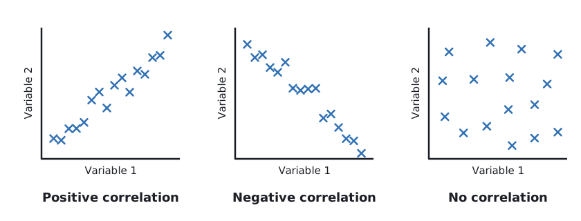
- Use a scatter diagram to identify a correlation between two variables. A positive correlation means one variable increases as the other increases. A negative correlation means one decreases as the other increases. No pattern means no correlation.
Sampling
It is not possible to test everyone, so scientists collect data from a sample. In epidemiological studies, a larger sample that is representative of the population gives more reliable data.
Disease incidence
In a town of 40 000 people, 60 new cases of a disease were recorded in one year. Calculate the number of new cases per 100 000 people.
- Divide the number of cases by the population: 60 ÷ 40 000 = 0.0015.
- Multiply by 100 000.
Answer: 150 cases per 100 000 people
 Tap to enlarge
Risk factors and causesSpec 4.2.2.6
Risk factors are linked to an increased rate of a disease. They can be:
- aspects of a person's lifestyle
- substances in the person's body or environment.
A causal mechanism has been proven for some risk factors, but not for others. A correlation between a risk factor and a disease does not prove that the risk factor causes the disease. A causal mechanism explains how the risk factor leads to the disease.
Many diseases are caused by the interaction of a number of factors. For example, CHD is affected by diet, smoking and how much exercise a person takes, as well as other factors.
The effect of lifestyle on diseaseSpec 4.2.2.6
You need to be able to explain the effect of lifestyle factors including diet, alcohol and smoking on the incidence of non-communicable diseases.
| Risk factor | Effect |
|---|---|
| Diet, smoking and exercise: cardiovascular disease | A diet high in saturated fat can raise blood cholesterol, which leads to fatty deposits in the arteries. Chemicals in tobacco smoke damage the artery lining and raise blood pressure, and carbon monoxide reduces the oxygen the blood can carry. Regular exercise reduces the risk |
| Obesity: Type 2 diabetes | Obesity is a risk factor for Type 2 diabetes |
| Alcohol: liver and brain | Drinking too much alcohol over a long time damages the liver. Alcohol also affects brain function, for example slowing reactions, and long-term heavy drinking can damage the brain |
| Smoking: lung disease and lung cancer | Smoking damages the lungs and airways, causing lung disease. Tar in tobacco smoke contains chemicals that cause lung cancer |
| Smoking and alcohol: unborn babies | Smoking in pregnancy can cause low birth weight and premature birth. Alcohol in pregnancy can damage the baby's development, including the brain |
| Carcinogens (including ionising radiation): cancer | A carcinogen is a substance or type of radiation that can cause cancer, such as chemicals in tobacco smoke. Ionising radiation, such as X-rays and gamma rays, can damage the DNA in cells |
Because lifestyles differ, the incidence of these diseases is different in different places. Diseases linked to diet and obesity, for example, are more common in places where people eat more high-energy food and take less exercise. This is true at local, national and global levels.
The cost of non-communicable diseasesSpec 4.2.2.6
You need to be able to discuss the human and financial cost of these diseases to an individual, a local community, a nation or globally.
| Level | Human cost | Financial cost |
|---|---|---|
| Individual | Pain, loss of independence, a shorter life | Lost income if the person cannot work. Costs of care and travel |
| Local community | Family and friends become carers. Loss of community members | Local businesses lose workers and customers |
| Nation | Many people with a reduced quality of life | Health services pay for treatment, drugs and operations. Lost working days reduce the national income |
| Global | Large numbers of early deaths and illnesses worldwide | Health care and lost work cost large amounts of money across the world |
Prevention through diet and lifestyle can reduce these costs, because many interventions would not be necessary if individuals reduced their risks.
Using data about risk factorsSpec 4.2.2.6
You need to interpret data about risk factors for specified diseases. The same skills apply as for incidence data.
- Translate information between graphical and numerical forms.
- Extract and interpret information from charts, graphs and tables.
- Use a scatter diagram to identify a correlation between a risk factor and a disease.
- Understand sampling: the sample should be large and representative for the conclusion to be reliable.
Describe what the data show first, then say whether there is a correlation. Then say whether the data prove a cause. A correlation alone does not. You also need a causal mechanism, or evidence that other factors were controlled.
CancerSpec 4.2.2.7
You need to be able to describe cancer as the result of changes in cells that lead to uncontrolled growth and division. These cells can form a lump called a tumour.
| Benign tumour | Malignant tumour | |
|---|---|---|
| Cells | Growths of abnormal cells | Cancer cells |
| Where they are | Contained in one area, usually within a membrane | Invade neighbouring tissues |
| Spread | Do not invade other parts of the body | Spread to different parts of the body in the blood, where they form secondary tumours |
| Cancer? | No | Yes |
Scientists have identified lifestyle risk factors for various types of cancer, such as smoking, obesity and drinking alcohol. There are also genetic risk factors for some cancers. Viruses living in cells can also trigger some cancers.
Quick check
Which enzyme breaks down proteins, and what are the products?
Show answer
Protease. It breaks proteins into amino acids.
Which ventricle pumps blood around the body?
Show answer
The left ventricle.
Name the three types of blood vessel.
Show answer
Arteries, veins and capillaries.
What does bile do?
Show answer
It neutralises stomach acid and emulsifies fat, increasing its surface area.
How do malignant tumours spread?
Show answer
Cells invade neighbouring tissues and spread in the blood to form secondary tumours.