

Eukaryotes and prokaryotesSpec 4.1.1.1
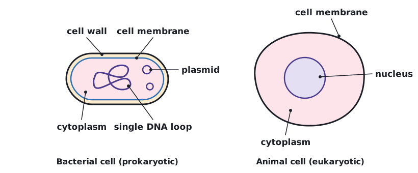
Plant and animal cells are eukaryotic cells. They have a cell membrane, cytoplasm and genetic material enclosed in a nucleus.
Bacterial cells are prokaryotic cells. They are much smaller in comparison. They have cytoplasm and a cell membrane surrounded by a cell wall. The genetic material is not enclosed in a nucleus. It is a single DNA loop, and there may be one or more small rings of DNA called plasmids.
| Eukaryotic cell (plant, animal) | Prokaryotic cell (bacterium) | |
|---|---|---|
| Size | Larger | Much smaller |
| Genetic material | Enclosed in a nucleus | Single DNA loop, not enclosed in a nucleus |
| Plasmids | No | One or more may be present |
| Cell membrane and cytoplasm | Yes | Yes |
| Cell wall | Plant and algal cells (made of cellulose); not animal cells | Yes |
 Tap to enlarge
Scale and size of cells
You need to understand the scale and size of cells and be able to make order of magnitude calculations. An order of magnitude is a power of ten. A cell that is 100 times wider than another is two orders of magnitude bigger, because 100 = 10².
| Prefix | Symbol | Standard form (compared with 1 m) | Example |
|---|---|---|---|
| centi | c | × 10⁻² | 1 cm = 0.01 m |
| milli | m | × 10⁻³ | 1 mm = 0.001 m |
| micro | µ | × 10⁻⁶ | 1 µm = 0.000 001 m |
| nano | n | × 10⁻⁹ | 1 nm = 0.000 000 001 m |
To change units: 1 mm = 1000 µm and 1 µm = 1000 nm. Standard form is a number between 1 and 10 multiplied by a power of ten, for example 0.000 004 m = 4 × 10⁻⁶ m.
Order of magnitude
A plant cell is 100 µm long. A bacterium is 1 µm long. How many orders of magnitude longer is the plant cell?
- Divide the larger length by the smaller: 100 ÷ 1 = 100.
- Write the answer as a power of ten: 100 = 10².
Answer: The plant cell is 10² (100) times longer, which is two orders of magnitude.
Standard form
A cell is 0.000 025 m wide. Write this in standard form, then in µm.
- Move the decimal point to give a number between 1 and 10: 2.5.
- Count the places moved: 5. Because the number is small, the power is negative: 2.5 × 10⁻⁵ m.
- To convert to µm, multiply metres by 1 000 000: 0.000 025 × 1 000 000 = 25.
Answer: 2.5 × 10⁻⁵ m, which is 25 µm.
Animal and plant cellsSpec 4.1.1.2
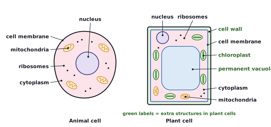
Most animal cells have: a nucleus, cytoplasm, a cell membrane, mitochondria and ribosomes. In addition, plant cells often have: chloroplasts and a permanent vacuole filled with cell sap. Plant and algal cells also have a cell wall made of cellulose, which strengthens the cell.
| Structure | Function | Animal cell | Plant cell | Bacterial cell |
|---|---|---|---|---|
| Nucleus | Contains the genetic material (DNA) and controls the activities of the cell | Yes | Yes | No |
| Cytoplasm | Where most chemical reactions happen | Yes | Yes | Yes |
| Cell membrane | Controls the movement of substances into and out of the cell | Yes | Yes | Yes |
| Mitochondria | Where aerobic respiration takes place, releasing energy for the cell | Yes | Yes | No |
| Ribosomes | Where proteins are made (protein synthesis) | Yes | Yes | Yes |
| Cell wall | Strengthens the cell; made of cellulose in plant and algal cells | No | Yes | Yes (not cellulose) |
| Chloroplasts | Contain chlorophyll, which absorbs light energy for photosynthesis | No | Yes (often) | No |
| Permanent vacuole | Filled with cell sap; helps keep the cell firm | No | Yes | No |
| Plasmid | Small ring of extra DNA | No | No | Possibly |
 Tap to enlarge
Estimating size and area
You should be able to use estimations to judge the relative size or area of sub-cellular structures. Round the measurements to easy numbers and compare them. Use an estimate when you only need to know roughly how much bigger one structure is than another, or to check that a calculated answer is sensible.
Estimating relative size
In a micrograph, a cell is 62 mm across and its nucleus is 19 mm across. Estimate how many times wider the cell is than the nucleus.
- Round the numbers: 62 mm is about 60 mm and 19 mm is about 20 mm.
- Divide: 60 ÷ 20 = 3.
Answer: The cell is roughly 3 times wider than the nucleus.
Required practical 1: using a light microscopeSpec 4.1.1.2
- Place a thin piece of tissue (for example onion epidermis, or cheek cells) on a clean microscope slide. Add a drop of water or a stain such as iodine solution.
- Lower a cover slip onto the specimen at an angle, using a mounting needle, so that no air bubbles are trapped.
- Clip the slide onto the stage. Select the lowest-power objective lens and turn the coarse focusing knob until the specimen is roughly in focus.
- Use the fine focusing knob to get a sharp image. Then switch to a higher-power objective lens and refocus with the fine focusing knob only.
- Draw what you see using a sharp pencil. Use clear, unbroken lines, no shading, and make the drawing large. Draw only a few cells accurately rather than many badly.
- Label the sub-cellular structures with straight label lines that do not cross. Add a title and the magnification.
To find the total magnification of a light microscope, multiply the magnification of the eyepiece lens by the magnification of the objective lens. The magnification of your drawing is found with the equation magnification = size of image ÷ size of real object, using the width of your drawing and the real width of the cell.
Safety: carry the microscope with two hands, handle glass slides and cover slips carefully, and take care with the stain, which can mark skin and clothes. Start with the lowest-power lens so that the objective lens does not hit the slide.
Cell specialisationSpec 4.1.1.3
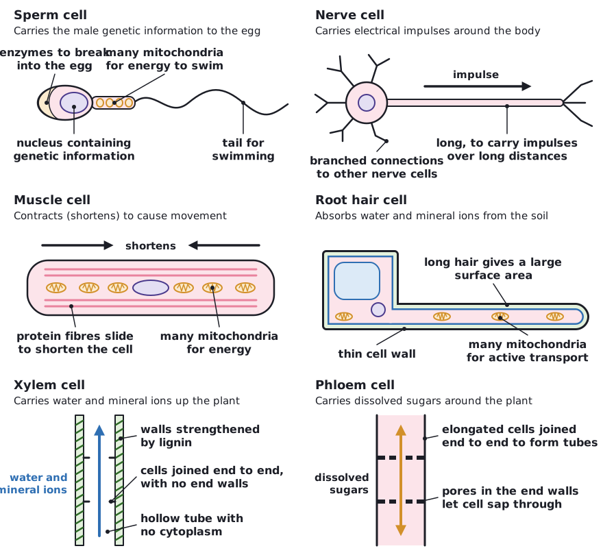
Cells may be specialised to carry out a particular function. You should be able to explain how the structure of different types of cell relate to their function in a tissue, an organ or organ system, or the whole organism, when you are given appropriate information.
| Cell | Function | How its structure helps |
|---|---|---|
| Sperm cell | Carries the male genetic information to the egg | Tail for swimming; many mitochondria to release the energy needed to swim; streamlined head; enzymes in the head to break into the egg; nucleus containing genetic information |
| Nerve cell | Carries electrical impulses around the body | Long (extended) so it can carry impulses over long distances; branched connections at the ends to connect with other nerve cells and organs |
| Muscle cell | Contracts (shortens) to cause movement | Contains protein fibres that can slide to make the cell shorten; many mitochondria to release energy for contraction; may store glycogen as an energy source |
| Root hair cell | Absorbs water and mineral ions from the soil | Long hair-like projection gives a large surface area; thin cell wall; many mitochondria to release energy for active transport of mineral ions |
| Xylem cell | Carries water and mineral ions up the plant | Joined end to end to form a hollow tube with no end walls and no cytoplasm; walls strengthened (by lignin) |
| Phloem cell | Carries dissolved sugars around the plant | Elongated cells joined end to end to form tubes; pores in the end walls let cell sap containing dissolved sugars move from one phloem cell to the next |
 Tap to enlarge
Cell differentiationSpec 4.1.1.4
As an organism develops, cells differentiate to form different types of cells. As a cell differentiates it acquires different sub-cellular structures to enable it to carry out a certain function. It has become a specialised cell.
- Most types of animal cell differentiate at an early stage.
- Many types of plant cell retain the ability to differentiate throughout life.
- In mature animals, cell division is mainly restricted to repair and replacement.
Differentiation is important because it allows cells to become specialised, with the sub-cellular structures they need for a particular job. A multicellular organism made of specialised cells can carry out many different functions efficiently, as the specialised cells work together in tissues, organs and organ systems.
MicroscopySpec 4.1.1.5
Microscopy techniques have developed over time. The first microscopes used light and lenses to magnify a specimen, and these light microscopes were improved with better lenses and the use of stains. Later, electron microscopes were developed. These use electrons instead of light.
An electron microscope has much higher magnification and resolving power than a light microscope. This means that it can be used to study cells in much finer detail. This has enabled biologists to see and understand many more sub-cellular structures.
| Light microscope | Electron microscope | |
|---|---|---|
| Magnification | Lower | Much higher |
| Resolving power (resolution) | Lower | Much higher |
| Detail seen | Cells and larger sub-cellular structures such as the nucleus | Much finer detail, including the internal structure of mitochondria and chloroplasts, and ribosomes |
- Magnification
- How many times larger the image is than the real object.
- Resolving power
- The ability to distinguish between two points that are close together as separate points. Higher resolving power gives a sharper, more detailed image.
Magnification calculations
Rearranged: real size = image size ÷ magnification, and image size = real size × magnification. Make sure the image and real object are in the same units before you divide, then convert at the end. Give the answer in standard form if appropriate.
Calculating magnification
A drawing of a cell is 45 mm wide. The real cell is 0.15 mm wide. Calculate the magnification.
- Both sizes are in mm, so no conversion is needed.
- magnification = 45 ÷ 0.15 = 300.
Answer: × 300
Calculating real size
A cell appears 12 mm wide in an image magnified × 400. Calculate the real width in µm.
- real size = image size ÷ magnification = 12 ÷ 400 = 0.03 mm.
- Convert to µm: 0.03 × 1000 = 30 µm.
Answer: 30 µm
Culturing microorganismsSpec 4.1.1.6Triple only
Bacteria multiply by simple cell division called binary fission, as often as once every 20 minutes if they have enough nutrients and a suitable temperature.
Bacteria can be grown in a nutrient broth solution or as colonies on an agar gel plate. Uncontaminated cultures of microorganisms are required for investigating the action of disinfectants and antibiotics, so that the only microorganisms present are the ones being tested.
Preparing an uncontaminated culture using aseptic technique
- Sterilise the Petri dish and the agar culture medium before use (for example by heating), to kill any unwanted microorganisms.
- Sterilise the inoculating loop by passing it through a flame.
- Use the sterile loop to transfer the microorganisms to the agar. Keep the lid of the Petri dish open for as short a time as possible.
- Secure the lid of the Petri dish with adhesive tape and store it upside down.
- Incubate the plate at 25 °C.
| Step | Reason |
|---|---|
| Sterilise Petri dishes and culture media | Kills unwanted microorganisms that could contaminate the culture |
| Pass the inoculating loop through a flame | Kills microorganisms already on the loop, so that only the intended microorganisms are transferred |
| Secure the lid with adhesive tape | Stops microorganisms from the air getting in, and stops the cultured microorganisms escaping |
| Store the dish upside down | Condensation drips onto the lid rather than onto the agar surface |
| Incubate at 25 °C in school laboratories | Reduces the chance of growing pathogens that are harmful to humans, which grow best at higher temperatures such as body temperature |
Culture calculationsSpec 4.1.1.6Triple only
Number of bacteria after a certain time
You should be able to calculate the number of bacteria in a population after a certain time if you are given the mean division time. Each division doubles the population.
- Work out the number of divisions: total time ÷ mean division time (use the same units).
- Number of bacteria = starting number × 2 to the power of the number of divisions.
Bacterial population
A single bacterium divides every 20 minutes. How many bacteria are there after 2 hours?
- 2 hours = 120 minutes. Number of divisions = 120 ÷ 20 = 6.
- Number of bacteria = 1 × 2⁶ = 64.
Answer: 64 bacteria
Standard form
A single bacterium divides every 20 minutes. How many bacteria are there after 8 hours? Give your answer in standard form to 2 significant figures.
- 8 hours = 480 minutes. Number of divisions = 480 ÷ 20 = 24.
- Number of bacteria = 2²⁴ = 16 777 216.
- Move the decimal point 7 places: 1.6777216 × 10⁷. To 2 significant figures: 1.7 × 10⁷.
Answer: 1.7 × 10⁷ bacteria
Areas of colonies and clear areas
You should be able to calculate the cross-sectional area of colonies, or of clear areas around colonies, using πr². The radius is half of the diameter.
Area of a clear zone
The clear zone around an antibiotic disc has a diameter of 14 mm. Calculate its cross-sectional area. Use π = 3.14.
- Radius = 14 ÷ 2 = 7 mm.
- Area = π × r² = 3.14 × 7 × 7 = 3.14 × 49 = 153.86 mm².
Answer: About 154 mm²
Required practical 2: antiseptics and antibioticsSpec 4.1.1.6Triple only
- Using aseptic technique, spread a sample of bacteria evenly over the surface of a sterile agar plate to make a bacterial 'lawn'.
- Using sterile forceps, place sterile paper discs soaked in different antiseptics or antibiotics (or different concentrations of one) onto the agar. Include a control disc soaked in sterile water.
- Secure the lid with adhesive tape, label the base, store the plate upside down and incubate at 25 °C for about two days.
- Measure the diameter of the clear area (zone of inhibition) around each disc and calculate its area using πr².
Where the antiseptic or antibiotic has killed or stopped the growth of the bacteria, there is a clear zone around the disc. The larger the zone of inhibition, the more effective the substance is against that bacterium. The control disc should have no clear zone, which shows that the water itself does not kill the bacteria.
- Independent variable: the type or concentration of antiseptic or antibiotic.
- Dependent variable: the size (diameter or area) of the zone of inhibition.
- Control variables: type and amount of bacteria, type and depth of agar, size of disc, volume of solution on the disc, temperature, incubation time.
Safety: the cultures may contain harmful microorganisms. Use aseptic technique, wash hands, keep the plate taped shut, never open it after incubation, and disinfect the work area and dispose of plates safely.
Quick check
How is the genetic material of a bacterial cell arranged?
Show answer
As a single DNA loop that is not enclosed in a nucleus. There may also be plasmids.
Which sub-cellular structure is the site of aerobic respiration?
Show answer
Mitochondria.
What is the formula for magnification?
Show answer
Magnification = size of image ÷ size of real object.
Why does an electron microscope show more detail than a light microscope?
Show answer
It has much higher magnification and resolving power.
Triple only: why are inoculating loops passed through a flame?
Show answer
To sterilise them and kill unwanted microorganisms.